
May 23
Discussions, inspiration, and a shared outlook on what comes next. 🤝
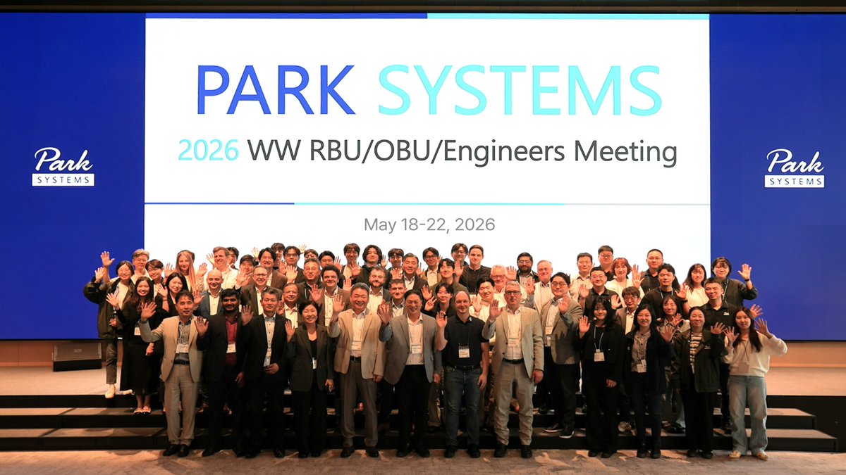
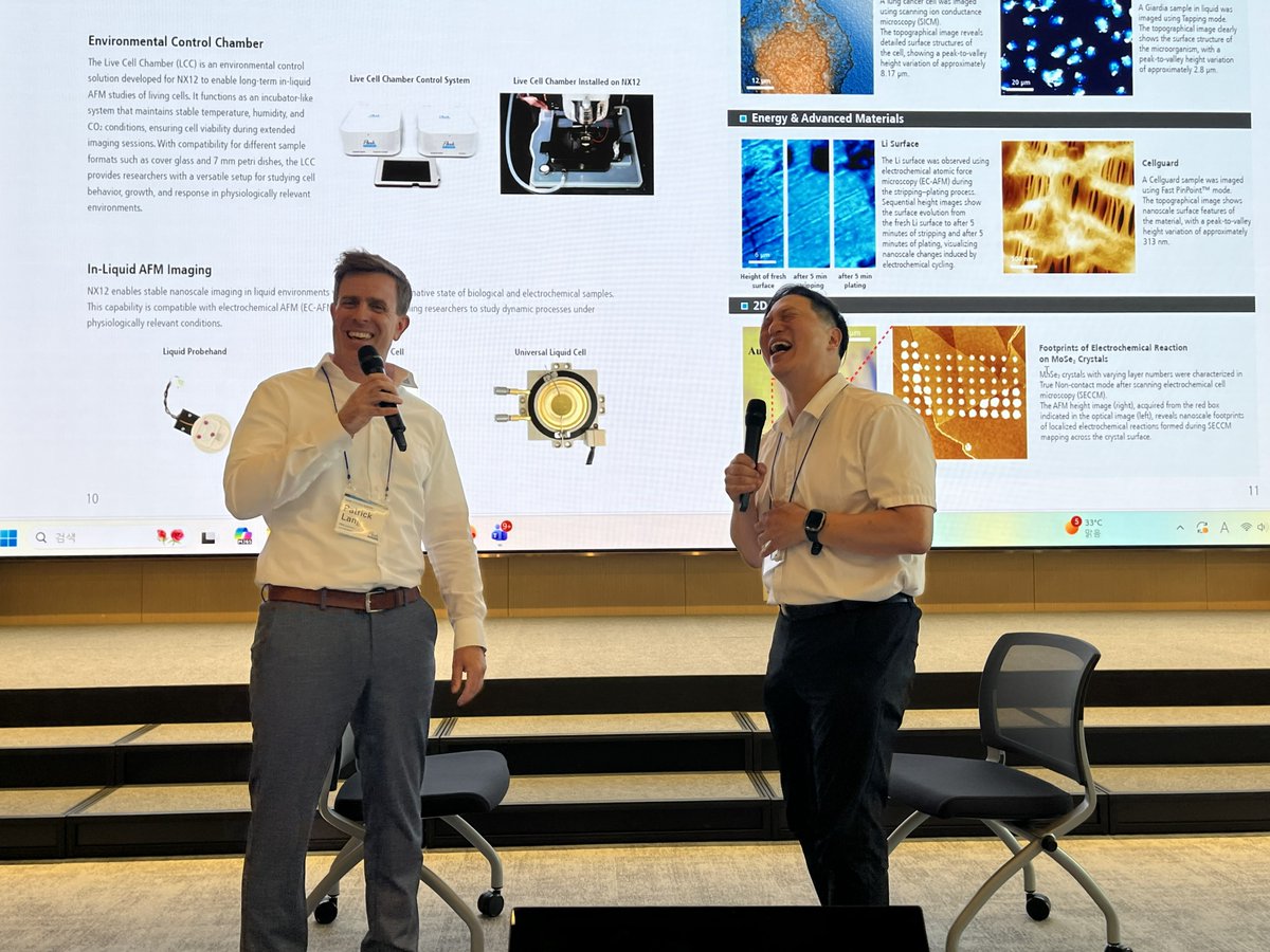
This week’s 𝗦𝗮𝗹𝗲𝘀 𝗮𝗻𝗱 𝗘𝗻𝗴𝗶𝗻𝗲𝗲𝗿𝗶𝗻𝗴 𝗠𝗲𝗲𝘁𝗶𝗻𝗴 𝗮𝘁 𝗣𝗮𝗿𝗸 𝗦𝘆𝘀𝘁𝗲𝗺𝘀 𝗛𝗤 in Korea brought our regional teams together to exchange ideas, learn from one another, and connect customer needs with technology development.
A strong team. Meaningful conversations. A clear direction forward. 🚀
#ParkSystems #Nanometrology #AdvancedMicroscopy #Teamwork


63

Apr 7
⏰ Did you miss the application deadline for #EMBLSuperResolution? 😱
We have good news for you: we have extended the deadline for the EMBL Course 'Time-resolved STED nanoscopy in life sciences' until 17 April 👉 s.embl.org/let26-01-x
Join us in Heidelberg to learn how to utilise STED microscopy for your biological questions. Explore how to acquire high-quality nanoscopy data on state-of-the-art STED microscopes 🔬
#STED #STEDMicroscopy #SuperResolution #Nanoscopy #AdvancedMicroscopy #FluorescenceMicroscopy #LifeSciences #CellBiology #ScientificTraining #MicroscopyCourse #EMBL #LeicaMicrosystems
2
4
947

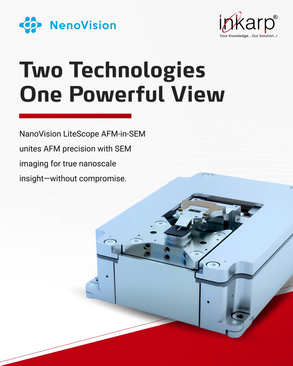
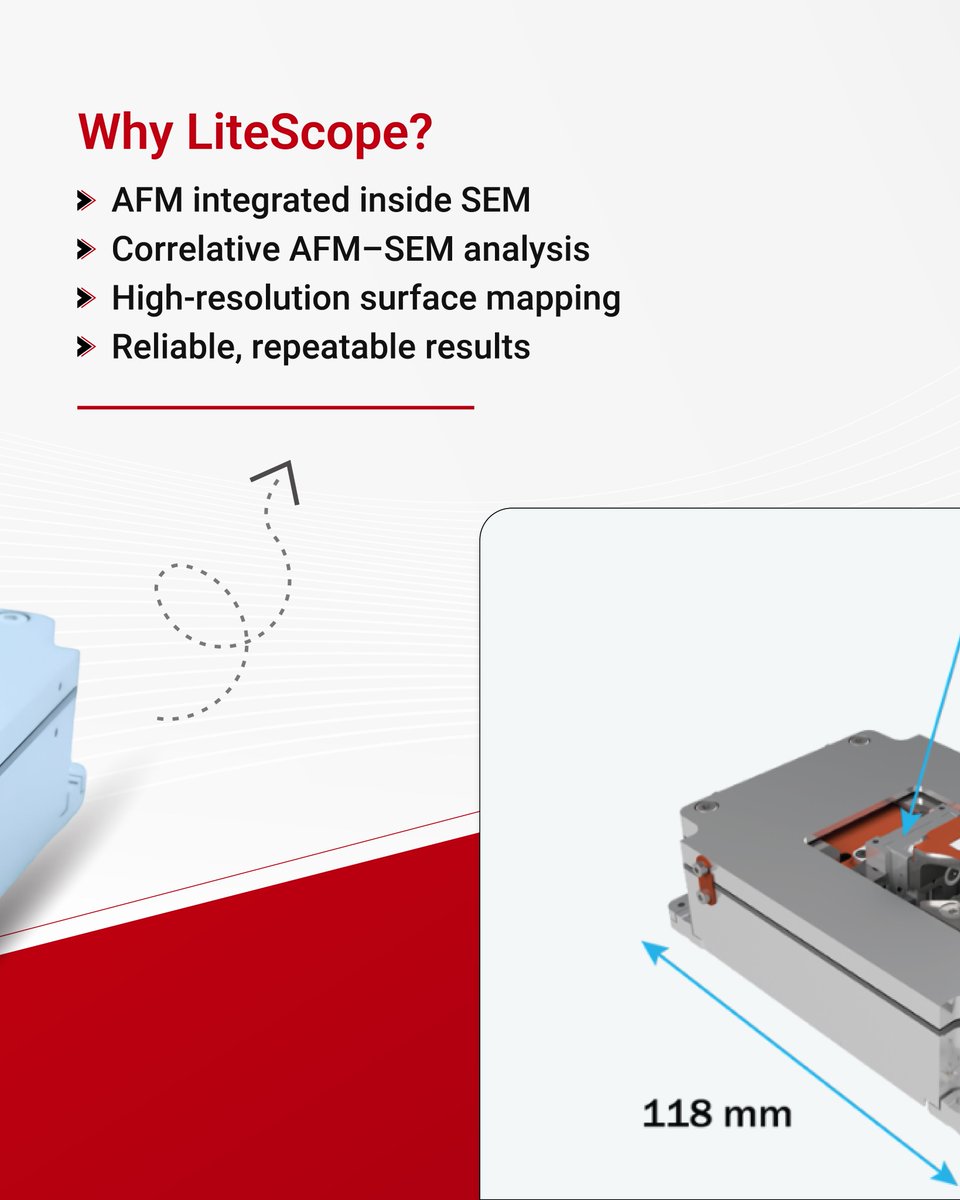
LiteScope AFM-in-SEM enables true correlative AFM SEM in one workflow.
Get Quote:
info@inkarp.co.in
inkarp.co.in/verticals/lifes…
#LiteScope #AFMinSEM #AdvancedMicroscopy


16

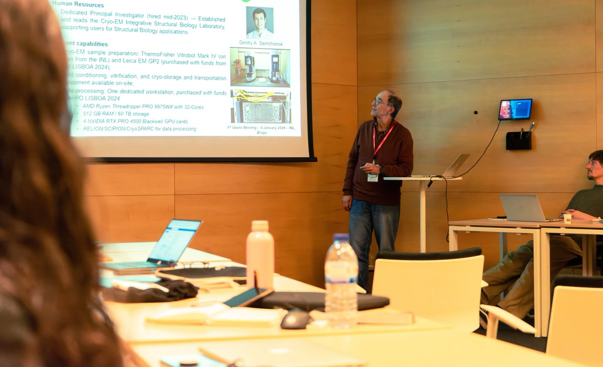
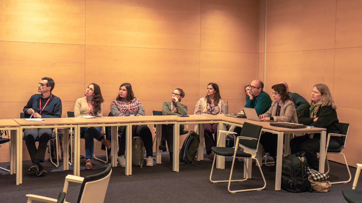
Advanced microscopy drives breakthroughs in areas like neurodegenerative disease research!
Scientists from INL, iBET, ITQB, CCMAR & FCT-UNL joined forces at the CryoEM-PT Users Meeting to define next steps for collaboration.
#CryoEM #AdvancedMicroscopy #INLnano

4
208

FLIM imaging, made faster, more precise, and more robust through the development of a photon-counting system for live visualization of cellular processes.
We are proud to participate in this!
#FLIM #AdvancedMicroscopy
Watch the interview (DE):
ost.ch/en/details/news/quant…
1
2
32

5 Dec 2025
🔔A tiny citrus pest is hiding a biological mystery never seen before🔔
A strange ribosome-filled tube inside a citrus-pest symbiont may rewrite bacterial biology and inspire new pest-control solutions.
Summary:
Scientists have discovered a strange tubular structure inside Profftella, a symbiotic bacterium in the Asian citrus psyllid. These long, helical tubes, filled with ribosomes, show a complexity not typically found in bacteria. The discovery reshapes ideas about bacterial evolution and internal architecture. It may also help create targeted methods to control a major global citrus pest.
Source: Toyohashi University of Technology (TUT)
👉 A multinational group of scientists has identified a previously unknown tubular structure within Profftella, a symbiotic bacterium linked to a major pest that affects citrus crops around the world. The finding was made possible through advanced microscopy techniques and could influence future approaches to pest management as well as research on how complex biological features evolve.
👉 A collaborative team from Pusan National University, the National Institute for Physiological Sciences, Kobe University, and Toyohashi University of Technology reported the discovery of this unusual tube-like structure within Candidatus Profftella armatura, which lives inside the Asian citrus psyllid (Diaphorina citri). Their results appear in Npj Imaging.
#Microbiology #BacterialEvolution #CellBiology #Symbiosis #InsectMicrobiome #PestManagement #CitrusGreening #BiologicalDiscovery #AdvancedMicroscopy #StructuralBiology #GenomicScience #EvolutionaryBiology #MicrobialEcology #ScientificResearch #BioInnovation #BiotechNews #LifeSciences #MolecularBiology #MicrobialStructures #NPJImaging #ScienceUpdate #ResearchBreakthrough #NewFindings #ScienceNews #LabDiscovery #BiologyFacts #GeneticsResearch #MicrobeWorld #BacteriaStudy #ScienceExplained #STEMNews #NatureResearch #BioResearch #SciComm
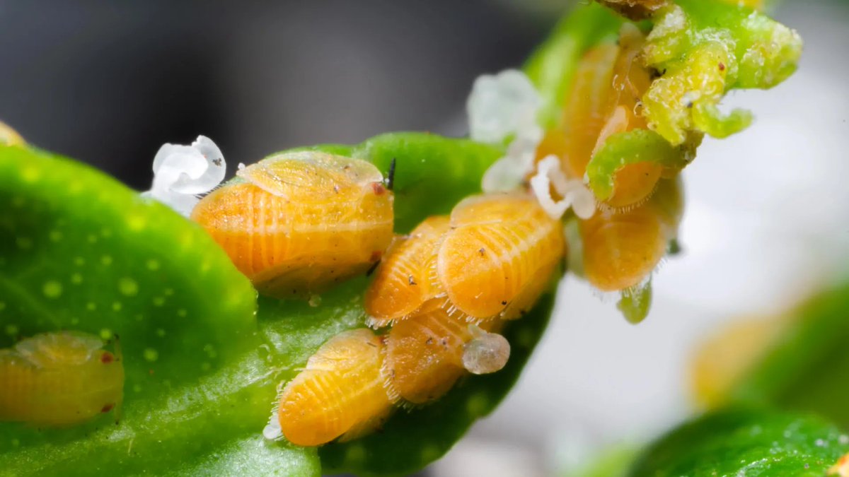
ALT A newly identified tubular organelle inside Profftella reveals unexpected complexity in a bacterium tied to the citrus-destroying psyllid. The finding could lead to novel biological insights and innovative pest-control approaches. Credit: Shutterstock
58

22 Nov 2025
How can we detect single photons for ultra-fast imaging?
Prof. Jörg Enderlein explains how SPAD arrays (single-photon avalanche diodes) detect individual photons and convert them into electric signals, enabling rapid, high-precision scanning that revolutionizes modern microscopy. These arrays are particularly powerful for applications like confocal microscopy, opening doors to previously impossible imaging speeds.
🎥 Watch Episode #21 with Prof. Jörg Enderlein from @uniGoettingen to explore how #SPADarrays, #PhotonDetection, and #AdvancedMicroscopy are enabling ultra-fast imaging and transforming #Biophysics research.
Neuroscience and Beyond: Episode Highlights
We hosted Prof. Jörg Enderlein, who shared how quantum principles intersect with modern microscopy. He explained key milestones in imaging, the link between spectroscopy and microscopy, and revolutionary tools like Airyscan and SOFI. The discussion highlighted how these innovations are driving breakthroughs in biophysics and expanding the limits of #microscopy.
#HighSpeedImaging #QuantumBiophysics #ConfocalMicroscopy #ImagingTechnology
Watch the full episode on this link: youtube.com/watch?v=n2e2kcrG…
Find us on social media: linktr.ee/neurosciencebeyond
1
3
97

9 May 2025
The #AQLM short course is in full swing at #MBL! Our high-quality scientific-grade CMOS cameras are proving to be invaluable assets in enhancing the imaging capabilities of advanced microscopy systems: ow.ly/e4t350VOWja
#Microscopy #CellBiology #AdvancedMicroscopy


1
92

4 Apr 2025
The 30th International Workshop on “Single Molecule Spectroscopy and Super-resolution Microscopy”, takes place on Sept 23–26, 2025, in Berlin.
❗The abstract due is April 16❗
More information:
➡️ow.ly/cZsc50VtKn8
#Event #Worksop #AdvancedMicroscopy #Imaging #Microscopy
71

25 Mar 2025
Assistant or Associate Professor in Electron Microscopy of Earth Materials (0.8 - 1.0 FTE) - Utrecht, Netherlands - earthworks-jobs.com/geoscien… #jobs #academicjobs #solidEarthSciences #Naturalsciences #advancedmicroscopy #ElectronMicroscopy
638


22 Jan 2025
Organized in collaboration with ZEISS, this training offered professionals the opportunity to explore the latest developments and applications in the field.
#MedipolSABİTA #ZEISS #MicroscopyTraining #ConfocalMicroscopy #SuperResolution #ScientificTraining #AdvancedMicroscopy
70

23 Dec 2024
Dr. Max's commitment to precision and patient care shines through his use of a high-definition wall-mounted microscope during surgery. This cutting-edge technology offers unparalleled visualization, allowing for enhanced detail and clarity in the surgical field.
The wall-mounted design provides superior stability and maneuverability, enabling Dr. Max to maintain optimal positioning throughout procedures.
The high-definition imaging not only improves surgical accuracy but also reduces eye strain and fatigue, allowing Dr. Max to maintain focus and perform at his best for extended periods.
This advanced microscope system also facilitates better collaboration among the surgical team, as the high-resolution images can be shared on large screens, enabling everyone in the operating room to follow the procedure closely.
By embracing this technology, Dr. Max demonstrates his dedication to delivering the highest standard of care, ensuring the best possible outcomes for his patients. The improved ergonomics of the wall-mounted system also contribute to Dr. Max's longevity in his profession, allowing him to continue providing exceptional surgical care for years to come.
#SurgicalExcellence #AdvancedMicroscopy #PrecisionSurgery #MedicalInnovation #PatientCare #SurgicalTechnology #HealthcareAdvancement #MedicalExpertise #SurgicalAccuracy #ModernMedicine
24

14 Nov 2024
With low-cost fabrication, this could revolutionise IVF labs, improving embryo selection and reproductive outcomes! ✨
🔗Find out more : tr.ee/MwoChZlzPy
#metabolicimaging #lightsheetmicroscopy #noninvasiveimaging #advancedmicroscopy #proofofconcept
57

12 Nov 2024
New arrivals in surgical precision and reliability—crafted for efficiency and control. Trusted by professionals worldwide. Discover more at angelusmedical.com.
#sampleanalysis #advancedmicroscopy #medicalequipments #medicalinstruments #healthclinic #equipment #instruments
8

10 Sep 2024
Nanoscale Optical Metamaterial Discoveries Gaining Speed
zurl.co/V4My
#NanoscaleOpticalMetamaterial #AdvancedMicroscopy #Spectrscopic #Interferometric #SumFrequencyImaging #RPMCLasers #lasers
13

27 Jun 2024
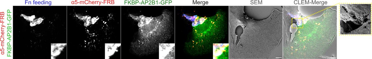
🔍With correlative light and electron microscopy (CLEM), we saw fibronectin aggregates getting internalized. This proves our hot-wired endocytosis works and modulates the ECM as well! #CLEM #AdvancedMicroscopy
1
1
126

18 May 2024
ポルトガルの研究者が画像解析AIプラットフォームを開発し、世界的な研究を促進
#DL4MicEverywhere #AIforMicroscopy #OpenScience #AdvancedMicroscopy
prompthub.info/5614/
1
2
36

6 May 2024
🔬Our Advanced Microscopy team brings together varied expertise to provide SME, academic and industrial partners with state-of-art imaging solutions.
Get in touch if you think we can help: hubs.li/Q02v_3L30
#AdvancedMicroscopy #LifeSciences #DrugDiscovery
1
250