Chidambara .ML. retweeted

Jun 12
Our #preprint list is now up on FocalPlane. This week we focus on the latest tools in bioimage analysis and include a couple of preprints that simulate microscopy data. Check them out and, as always, let us know if you have any recommendations.
focalplane.biologists.com/20…
1
5
19
2,043

[PUBLI] The challenge of GloBIAS 🖥️
In an article published in Nature Methods, the GloBIAS community presents the origins of the society, its missions and the actions to support the development of bioimage analysis worldwide.
To read our full article👉 france-bioimaging.org/announ…

2
4
148

Jun 4
The sixth session of BIOINFOCONGRESS VIII is now available.
Dr. Aybüke Küpçü Yoldaş’s talk titled
“FAIR Bioimage Data: What EMBL-EBI Provides?”
along with the Q&A session is now on the Bioinforange YouTube channel.
Bring your scientific curiosity and your headphones.
YouTube: Bioinforange
#bioinforange #bioinfocongressviii #fairdata #bioimaging #bioinformatics
3
100

Also in Issue 10:
- Research Highlights on ring canals in Drosophila egg chambers and mechanotransduction in Merkel cells
- Practical statistics for bioimage analysis
- Amoeboid cancer cells at a glance
journals.biologists.com/jcs/…

ALT JCS poster on amoeboid cancer cells at a glance
4
240

May 30
Yeah you would have to request more :(. I need to set up an integration with bioimage archive…
2
1,537

#HappyFluorescenceFriday #microscopycommunity- Join us June 4 for “2D Image Analysis Workflow with QuPath” featuring Lhorane Lobjois and learn practical workflows and open-source tools for bioimage analysis using QuPath.
Learn more: buff.ly/8pzsrI3

2
3
151
[REPLAY] BioImage Cloud webinar now available on YouTube ⏮️
We were delighted to host Guillaume Gay, who introduced BioImage Cloud, the data management solution developed by the FBI.data team.
Watch the replay 👉youtube.com/watch?v=-2hAZlOq…

2
2
133
BioImage Cloud is officially launched! 📢
The data management tool developed by the FBI.data team is now available & will be deployed across FBI facilities.
👉Read our article to access all the resources created to help you get started: bit.ly/4v0u28X
2
3
212

May 6
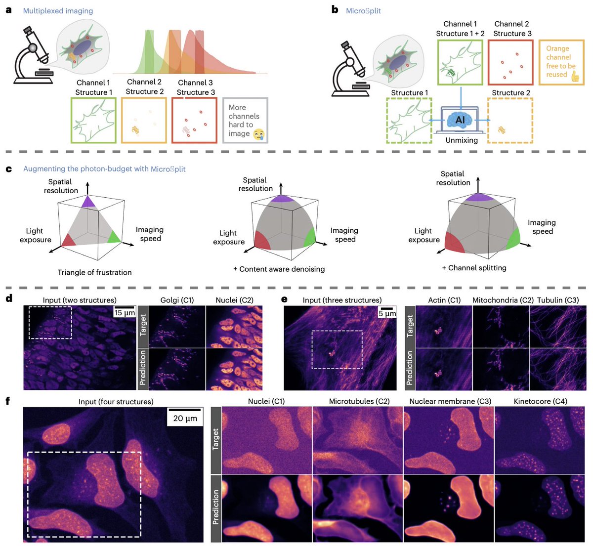
Imaging more cellular structures than your microscope allows with variational encoder-decoders
Fluorescence microscopy is bound by a stubborn trade-off triangle. You want spatial resolution, imaging speed, and low light exposure, but you can only really push two at a time. The number of fluorophores you can use simultaneously is also capped by spectral overlap between dyes, and every extra channel eats into a finite photon budget. For live-cell imaging, where phototoxicity matters, this is a hard ceiling on what you can see.
Ashesh Ashesh and coauthors flip the problem: instead of separating structures optically, label and image multiple structures into a single fluorescent channel, then unmix them computationally.
Their method, MicroSplit, is built on Variational Splitting Encoder-Decoder networks, a hierarchical variational architecture that learns a posterior over plausible unmixed solutions rather than a single point prediction. It jointly performs supervised channel splitting and unsupervised denoising using a learned noise model, so the network can be trained on noisy targets and still output denoised predictions for each structure.
Across 30 tasks spanning ten datasets, MicroSplit cleanly separates up to four superimposed structures (nuclei, microtubules, nuclear membrane, kinetochores) from a single channel. Average PSNR sits at 32.5 and microMS-SSIM at 0.89, comfortably in the range used for downstream segmentation. Segmentation quality on unmixed images stays within inter-observer variability of three bioimage analysts working on conventionally multiplexed data.
A nice feature is that the variational network gives calibrated uncertainty estimates. By sampling 50 posterior solutions per input and computing inter-sample variance, the method produces a pixel-wise error map that correlates linearly with true error, addressing a persistent pain point in AI for bioimaging: knowing where to trust the model.
The authors also show MicroSplit can remove structured imaging artifacts (spurious puncta) by treating them as just another structure to unmix, and that the freed photon budget allows roughly a tenfold reduction in light exposure at comparable quality.
For drug discovery and biotechnology, this changes the cost structure of high-content screening and live-cell phenotyping. Imaging more targets per well with lower phototoxicity means longer time-lapse experiments and richer readouts without buying new optics. The calibrated uncertainty maps are especially relevant for regulated pipelines, where flagging unreliable regions matters more than squeezing out the last decimal of accuracy.
Paper: Ashesh et al., Nature Methods (2026) — CC BY 4.0 | doi.org/10.1038/s41592-026-0…
13
56
5,086

🎉 A new Node joined #EuroBioImaging! The Smart Optical Microscopy (SOM) Madrid Node 🇪🇸 will provide cutting-edge services in advanced fluorescence microscopy, with a focus on #SmartMicroscopy, high-throughput & AI-powered bioimage analysis.
More⤵️
eurobioimaging.eu/news/welco…
2
6
275
Our latest preprint list is now up on FocalPlane. This week, bioimage analysis takes centre stage!
focalplane.biologists.com/20…
#microscopy #bioimageanalysis #preprints
1
5
633

Apr 25
GloBIAS: strengthening the foundations of bioimage analysis | Nature Methods nature.com/articles/s41592-0…
2
13
557

Apr 23
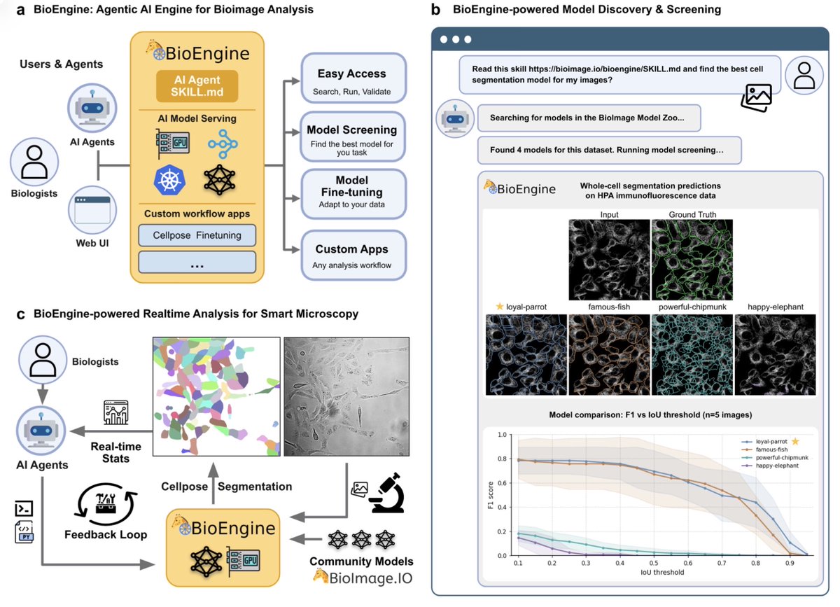
BioEngine: scalable execution and adaptation of bioimage AI through agent-readable interfaces biorxiv.org/content/10.64898…
1
4
8
448

Apr 23
Super excited to share my first PhD paper! 🎉 We built #BioEngine, a scalable bioimage AI platform where AI agents can screen, run, and fine-tune models. From a lab workstation to an institutional cluster, one-time setup.
📄 biorxiv.org/content/10.64898…
More in the thread 👇
1
2
6
269
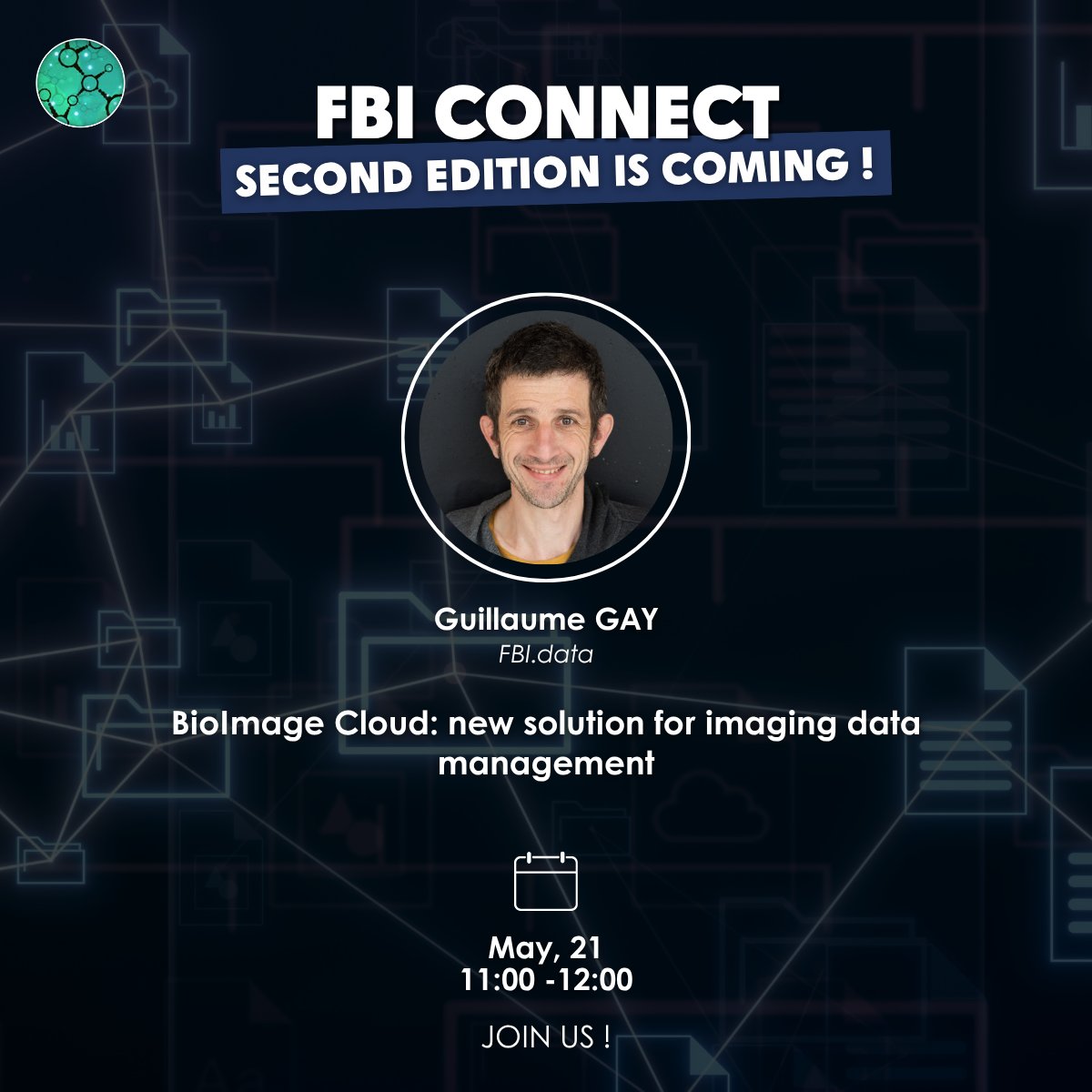
Working with large imaging datasets?
Join the next FBI Connect webinar to discover BioImage Cloud (BiClou), a solution for France-BioImaging facilities connecting OMERO & storage to streamline data management.
📅 May 21, 11:00 AM
🎤 G. Gay
🔗 bit.ly/4vtKJKO
1
3
4
260