
🔶The humanoid shaped structures on endoscopic examination represent colonic pseudopolyps (post-inflammatory polyps), which are non-neoplastic mucosal remnants resembling stick figures.
∆ Etiology: They arise during the healing phase of severe mucosal ulceration, where islands of regenerating granulation tissue form raised projections.
• This classic finding is heavily associated with Inflammatory Bowel Disease (IBD), particularly long standing Ulcerative Colitis.
∆ Clinical Management: They require periodic endoscopic surveillance and medical management of IBD, with polypectomy reserved only for symptomatic or obstructive lesions.
162

Peutz–Jeghers Syndrome (PJS)
1. Autosomal dominant disorder
Caused by mutation in STK11 (LKB1) tumor suppressor gene.
2. Mucocutaneous pigmentation
Dark blue-brown melanin spots on lips, oral mucosa, fingers, and perioral area.
3. Hamartomatous polyps
Multiple polyps in small intestine (especially jejunum), also stomach and colon.
4. Histology of polyps
Characteristic tree-like branching of smooth muscle within polyp.
5. Age of presentation
Usually childhood or adolescence (pigmentation often appears early).
6. Main clinical issue
Recurrent intestinal obstruction, intussusception, abdominal pain.
7. Bleeding risk
GI bleeding → anemia due to multiple polyps.
8. Cancer risk (very important)
Increased risk of multiple malignancies:
Pancreatic (high yield)
Colorectal
Gastric
Breast
Ovarian/testicular tumors
9. Diagnosis
Based on:
Typical pigmentation polyps
Family history
Endoscopy/biopsy confirmation
10. Management
Regular endoscopic surveillance
Polypectomy to prevent obstruction
Cancer screening lifelong.
6
314

🎤 Polypectomy / EMR (Colon)
@salahudinkhalid delivers an insightful session on Polypectomy and Endoscopic Mucosal Resection (EMR) in the Colon, sharing practical techniques and best practices for effective endoscopic management of colorectal lesions.#TECNA2026 #ScopeHealthUK


44

Jun 13
procedure was performed. During the colonoscopy, if doctors see a polyp — a small growth inside the colon — they usually remove it right away (polypectomy). In my case, a non-cancerous polyp in descending colon was removed via cold snare polypectomy (JNET 2A). The tissue was then
1
25

The image from the colonoscopy shows a smooth, rounded, pedunculated (stalk-like) mass projecting into the lumen of the colon. This is a classic presentation of a colonic polyp, specifically a pedunculated adenomatous polyp.
Why This Finding is Clinically Important
The detection of such polyps is the cornerstone of colorectal cancer prevention.
Precancerous Potential: Adenomatous polyps are considered neoplastic. While they are benign at the time of discovery, they possess the potential to transform into colorectal adenocarcinoma over time through the "adenoma-carcinoma sequence."
The "Adenoma-Carcinoma Sequence": This describes the genetic and histological progression from normal colonic epithelium to a small adenoma, to a large/advanced adenoma, and finally to invasive cancer.
Asymptomatic Nature: As noted in the post ("No symptoms. No pain."), these polyps are often silent. This is precisely why screening colonoscopy is critical; it allows for the identification and removal of these lesions before they ever have the chance to become malignant.
Clinical Management
When a mass like this is identified during a colonoscopy, the following steps are typically taken:
Polypectomy: This is both diagnostic and therapeutic. The polyp is removed during the procedure using a wire snare (often with electrocautery) to cut through the stalk.
Histopathological Examination: The removed polyp is sent to a pathologist. They determine the type (e.g., tubular adenoma, tubulovillous adenoma) and check for the presence of high-grade dysplasia or invasive cancer.
Surveillance Planning: Based on the size, number, and histological findings of the polyp, the gastroenterologist will determine the appropriate interval for the patient's next surveillance colonoscopy.
41

Jun 12
แทบไม่เคยเห็น polypectomy แล้ว bleed จน hct drop เลยคุณมา ทำไมต้องเกิดกับน้องนี่ด้วย แงง 😭
1
71

Six Parallel Masterclass Stations
Each station:
30 min focused lecture
90 min hands-on
Total: 120 min
1. Advanced Polypectomy & EMR
Lecture (30 min)
Lesion assessment (Paris classification, NICE/JNET)
Cold snare vs hot snare
EMR techniques
Prevention of post-polypectomy complications
Hands-on (90 min)
Cold snare polypectomy
Pedunculated polyp resection
EMR with submucosal injection
Specimen retrieval
2. Endoscopic Submucosal Dissection (ESD)
Lecture (30 min)
Indications and patient selection
Marking, injection, circumferential incision
Traction methods
Management of perforation and bleeding
Hands-on (90 min)
Knife handling
Mucosal incision
Submucosal dissection on ex-vivo models
3. Defect Closure & Perforation Management
Lecture (30 min)
Through-the-scope clips
Over-the-scope clips
Purse string closure
Endoscopic suturing overview
Closure algorithms
Hands-on (90 min)
Clip closure
Large defect closure techniques
OTSC deployment (if available)
Endoloop
Sutuart
4. Endoscopic Hemostasis
Lecture (30 min)
Non-variceal bleeding
Post-ESD/EMR bleeding
Mechanical, thermal, and topical therapies
Hemostasis algorithms
Hands-on (90 min)
Clip placement
Coagulation forceps
APC
Hemostatic powder application
5. Bariatric Endoscopy
Lecture (30 min)
Intragastric balloons
Endoscopic sleeve gastroplasty (ESG)
Revisional bariatric endoscopy
Patient selection
Hands-on (90 min)
Suturing platform demonstration
ESG simulation
Balloon placement/removal
6. EUS-Guided Tissue Acquisition (FNA/FNB)
Lecture (30 min)
Needle selection
FNA vs FNB
Sampling techniques
ROSE and specimen handling
Hands-on (90 min)
Scope positioning
Needle puncture techniques
Pancreatic mass and lymph node models


7
556

Jun 11
[JUNE 24th] Learn about safe #Polypectomy techniques & closure strategies.
➡️REGISTER from your mobile device => endoscopynowapp.app.link/XBI…
➡️Sign up on Zoom=> us06web.zoom.us/webinar/regi…
@bostonsci @bsc_endoscopy #Gastroenterology #GIEndoscopy #GIFellows #GITwitter #GINurse #TwitterMed

98

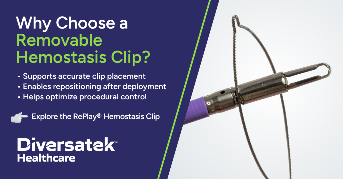
A removable hemostasis clip provides clinicians with real-time control & flexibility, enabling confident treatment.
Reposition, remove, & re-treat with the RePlay® Clip as needed. Use polypectomy snares for straightforward removal.
#GITwitter
Link: okt.to/MwUlRO
1
14
Yan Chu, MD retweeted

Tips for Colon Polypectomy
I would also add: have clips and injector needles available, know how to perform a submucosal cushion, do not perform polypectomy in dirty colon
28
71
3,179

Jun 10
Party petty pity purity potty pretty play portray posterity prosperity property properly paltry playboy panoply pantry purely polygamy polypectomy plutocracy probably probability penny patchy pithy poultry policy patchy pony puberty publicity poignancy pastry pornography poverty
43