
Passionate Scientist | Senior Lecturer | Vascular Biology | Bone & Bone Regeneration | Lab @MPI_Muenster | Steering Group MPG LeadNet | Enthusiastic Aikidoist
Joined June 2024
- Tweets 109
- Following 111
- Followers 84
- Likes 557
12 Photos and videos
Gabi Bixel retweeted

18 Oct 2025
In 1992 Peter Ratcliffe received this rejection letter from Nature.
His findings were not "a sufficient advance in our understanding".
27 years later he won the Nobel Prize for the same discovery.
Don't lose faith in the things you believe in.

173
1,875
7,726
728,565

The suggestions for the ECTS 2026 Programme are open!
Suggestions are welcomed for incorporation into the following sessions: Plenary Symposia, Clinical Concurrent Symposia, Basic Science Concurrent Symposia.
buff.ly/4b7bzig
5
7
629
Gabi Bixel retweeted

25 Sep 2025
Dr. @ralfhadams and team show that a small subpopulation of #endothelial cells inside the intestinal villi of the embryonic mouse gives rise to arterial endothelium in the intestinal wall but also in the distant mesenteric vasculature.
🆕 go.nature.com/3VBd1m0
1
6
30
1,077
Gabi Bixel retweeted

26 Sep 2025
Finally out! So happy to share my postdoctoral work with @ralfhadams and colleagues!
In this study, we uncover how arteries form in the embryonic gut and mesentery.
nature.com/articles/s41467-0…
6
14
46
2,427
Gabi Bixel retweeted

4 Oct 2025
We are hiring!! PhD positions available in my group at @UK_Muenster to study the role of inhibitory receptors in the regulation of inflammation! jobs-sf.ukmuenster.de/job/UK…
Please share and apply!
17
20
3,131
Gabi Bixel retweeted

10 Oct 2025
BM niche and vasculature work isn’t for the faint of heart. It takes serious technical precision.
9 Oct 2025

In a recent preprint at bioRxiv (Yang et al., doi.org/10.1101/2025.10.02.6…), a group of authors led by Dr Anjali Kusumbe challenges an article from my lab published in 2024 (Koh et al. 2024, PMID: 39537918). The new preprint refers to Extended Data Figures and Supplementary videos that are unfortunately not provided at bioRxiv. Nevertheless, the manuscript text and the 5 main figures plus a proposed model contain a couple of major issues that I will cover here.
1/
1
3
17
4,273
Gabi Bixel retweeted

10 Oct 2025
Thank you so much, Ruslan. I often wonder how we can improve the way we make research, publish, review and interact in science.
Sadly, not everyone seems to like open discussion

1
2
12
713
Gabi Bixel retweeted
10 Oct 2025
Much earlier than expected, it is time for another short thread on a recent preprint at bioRxiv (Yang et al., doi.org/10.1101/2025.10.02.6…) by a group of authors led by Dr Anjali Kusumbe.
Today, I want to cover some of the bioinformatics results presented in their preprint. As a reminder, based on the reanalysis of our scRNA-seq data from Koh et al. (PMID: 39537918), it is claimed in Fig. 1 and the accompanying text that lymphangiogenic markers are downregulated in aged skull samples.
We now had the chance to closely examine the relevant data, which has uncovered a couple of major issues.
1/4
2
9
45
9,265
Gabi Bixel retweeted

20 Sep 2025
Identification of lymphatic vessels in skull periosteum but not bone marrow reveals skull channel heterogeneity rupress.org/jem/article/222/… @JExpMed
13
57
4,016
Gabi Bixel retweeted

10 Sep 2025
We are delighted to share the review article regarding CSF draining lymphatics in health and disease: advances and controversies.
nature.com/articles/s44161-0…
1
16
51
4,014
Gabi Bixel retweeted
9 Oct 2025
In a recent preprint at bioRxiv (Yang et al., doi.org/10.1101/2025.10.02.6…), a group of authors led by Dr Anjali Kusumbe challenges an article from my lab published in 2024 (Koh et al. 2024, PMID: 39537918). The new preprint refers to Extended Data Figures and Supplementary videos that are unfortunately not provided at bioRxiv. Nevertheless, the manuscript text and the 5 main figures plus a proposed model contain a couple of major issues that I will cover here.
1/
5
30
93
42,681
Gabi Bixel retweeted
9 Oct 2025
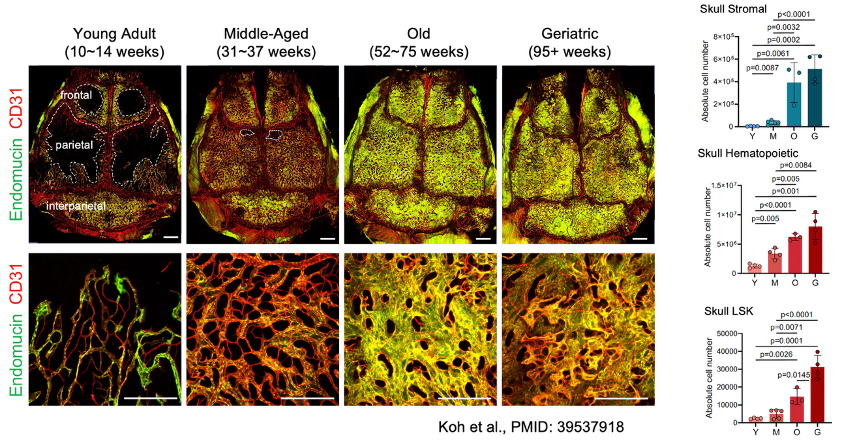
Apart from imaging, we provide an extensive set of flow cytometry data in a large (22 page) supplemental file accompanying the paper, which confirms the expansion of stromal, endothelial, total hematopoietic cells and various hematopoietic cell subsets during adulthood and aging but also in response to various challenges (such as pregnancy and stroke).
4/
1
5
16
3,675
Gabi Bixel retweeted
9 Oct 2025
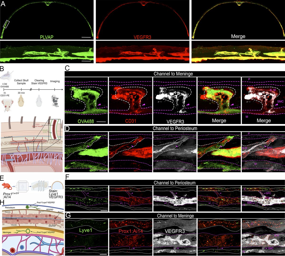
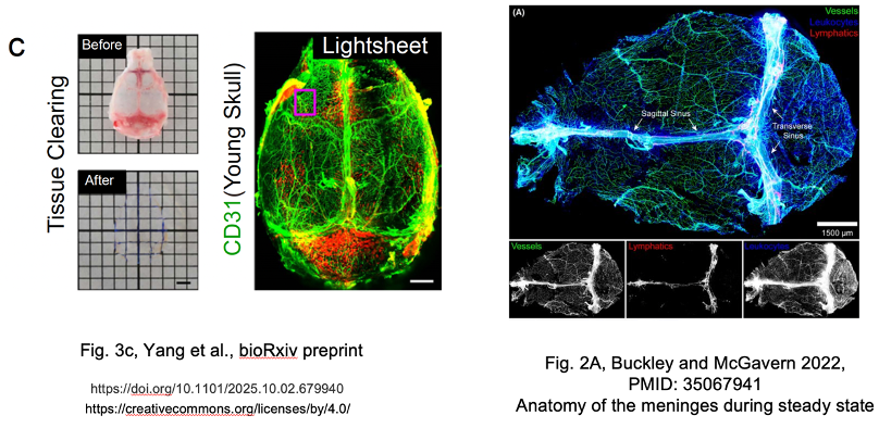
Things get more interesting and controversial in Figure 3c and d. Yang et al. use tissue clearing and light sheet microscopy to show the presence of a “continuous, dense vascular network throughout the skull frontal, parietal and interparietal regions in young mice”. The CD31 immunostaining in Fig. 3a, however, shows predominantly the meningeal vasculature, easily identifiable by hallmarks such as the sagittal and transverse sinuses consisting of large diameter blood and lymphatic vessels. Periosteal vessels might be also labeled, but it is not obvious that any sinusoidal vessels of the bone marrow are stained in this sample.
9/
1
6
17
3,180
Gabi Bixel retweeted
9 Oct 2025
I want to conclude here and thank all of you who have read this thread till the end. As I have not been able to cover all issues in the preprint, I am considering another detailed thread once the Extended Data becomes public.
I also hope that I have remained factual and sufficiently polite throughout my posts even though I perceive the language in the Yang et al. preprint as sometimes overly harsh and many claims unsubstantiated and wildly exaggerated.
Final full disclosure: I am not only the last author of Koh et al. 2024 (PMID: 39537918) but also the former postdoctoral supervisor of the two last authors of the Yang et al. preprint.
17/17
1
7
42
3,572
Gabi Bixel retweeted

🧩 "Lymphatic mysteries unzipped"
Our N&V @NatureCVR on the beautiful study of @hansschoofs @makinenlab uncovering the mechanics behind the oak-leaf shape of lymphatic ECs!Fun to work together with Gwen Randolph!
nature.com/articles/s44161-0…
Original paper: nature.com/articles/d41586-0…
1
7
35
1,664
Gabi Bixel retweeted

11 Apr 2025
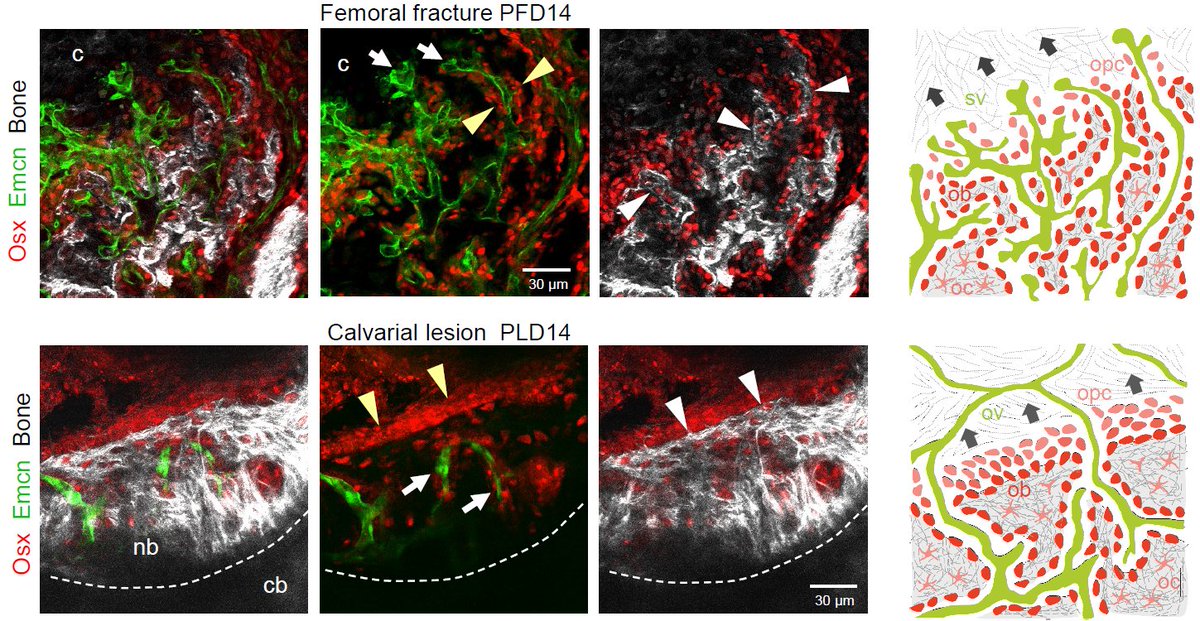
#FluorescenceFriday High-res confocal imaging showing Osx⁺ osteoprogenitors and angiogenesis during bone healing. Coupled or uncoupled 🫶, Either way, they seem happy to help.
#BoneHealing #Regeneration #imaging
3
10
68
3,455
Gabi Bixel retweeted

17 Mar 2025
Make a Break: Using spatial transcriptomics to study mechano-regulation of fracture healing
📷: Neashan Mathavan et al @eth_bone lab @ETH
in @ScienceAdvances
➡️: bpod.org.uk/archive/2025/3/1… with @DrJohnAnkers
5
18
1,279


Gabi Bixel retweeted

2 Jan 2025
The newly established Helmholtz Institute for Translational AngioCardioScience (HI-TAC) is looking for two Junior Research Group Leaders in the field of AngioCardioScience! Please feel free to reach out and help spread the word! Apply now: application.mdc-berlin.de
17
19
3,924
Gabi Bixel retweeted

11 Feb 2025
‘We must...fiercely protect the progress women in science have made.’ 🔬On #WomenInScienceDay, don’t miss this insightful interview with Suropriya Saha, Research Group Leader @mpids, on the legacy of #Physics Nobel laureate Maria Goeppert Mayer🌟 ▶️mpg.de/23712159/suropriya-sa…
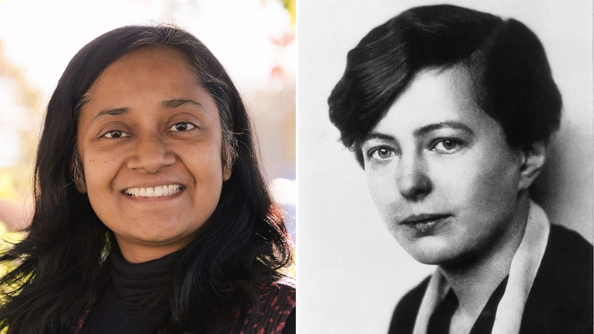
ALT Left: Suropriya Saha, Research Group Leader at the Max Planck Institute for Dynamics and Self-Organization (Credit: private) Right: Maria Goeppert Mayer (Credit: SUB Göttingen / Sammlung Voit)
1
15
43
4,492