
🤦🏻♂️🧢👓🏝🔬🦠🧫 Senior Scientist at UC San Diego department of Neurosciences
Joined February 2019
- Tweets 1,115
- Following 474
- Followers 497
- Likes 4,308
158 Photos and videos


Gerard Lambert 🏳️🌈 retweeted

14 Jul 2023
Excited to share our recent paper published in @ACSSynBio on our work developing genetically encoded bioluminescent neurotransmitter sensors for glutamate! pubs.acs.org/doi/10.1021/acs… with @GElegans1 @NathanShaner @LABrechaun @_theglowup and many others who contributed.
1
5
13
1,207

18 Feb 2023
HeLa cells are so awesome when stimulated with histamine 🥹This is bioluminescence imaged on our Nikon Widefield w/ @NathanShaner @_theglowup
1
1
39
4,267
Gerard Lambert 🏳️🌈 retweeted

28 Jan 2023
About 4 minutes of action from our best new intensiometric bioluminescent calcium indicator in HeLa cells exposed to l-histamine. Sorry for no time stamp or scale bar 🥸 Seeing ΔL/L of >30-fold in some cells 👀 w/@LABrechaun, contributions from @_theglowup, biorXiv soon🤞
3
12
86
12,992
28 Oct 2022
I hope Anya Salih sees this photo❤️ @SaliAnya remembering the fun times coral prospecting in Australia w/@NathanShaner
4
16 Sep 2022
Had fun imaging HEPG2 cells🙂 the bottom cell has a lot of personality
w/ @NathanShaner #FluorescenceFriday
1
8
Gerard Lambert 🏳️🌈 retweeted

12 Jul 2022
A great example of how important it is to get rid of #aberrations, to improve your #resolution, & to have a more sensitive #detection - True for telescopes as well as microscopes 🔬💪😀 - Well done, @NASAWebb & @NASA! #imagequality
11 Jul 2022

I had to make a before and after to really appreciate how good the James Webb Telescope really is.
1
6
27
9 Jul 2022
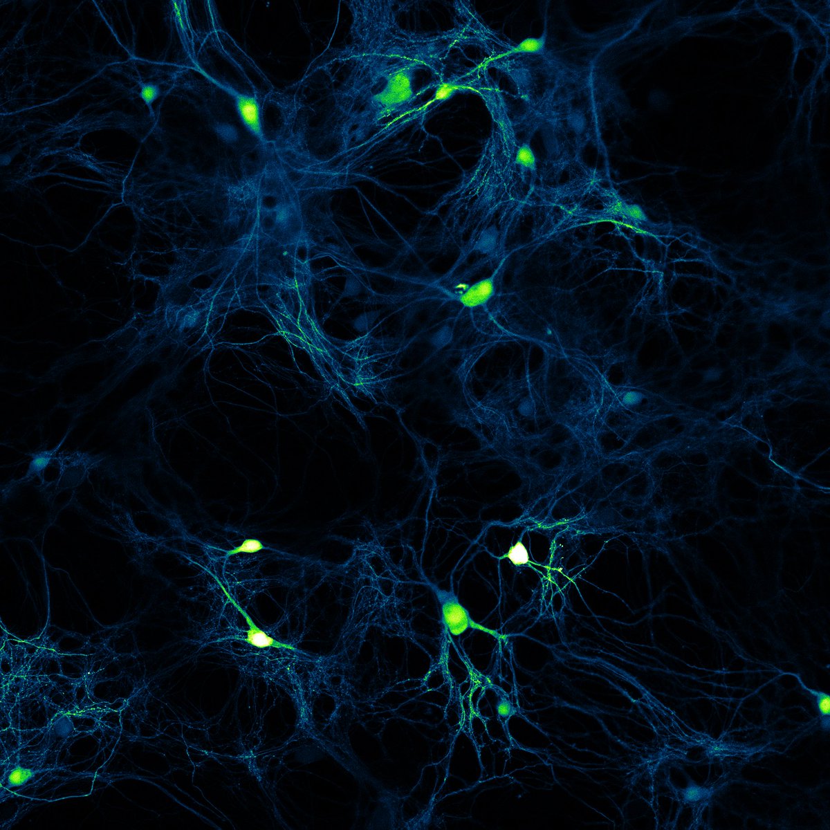
one of our summer student (not on Twitter) found this cool geometric actin structure in a U2OS cell 🤩
#FluorescenceFriday w/@NathanShaner
9
33
453
Gerard Lambert 🏳️🌈 retweeted

10 Jun 2022
Happy #FluorescenceFriday! Check out this time-lapse movie showing the extrusion of larval epidermal cells during Drosophila development. Adherens junctions are visualized by E-cadherin-GFP.
@Dev_journal publication: doi.org/10.1242/dev.179606
2
35
225
Gerard Lambert 🏳️🌈 retweeted

1 Jun 2022
Uri Manor @manorlaboratory & I are organizing a new imaging course! Join us at @salkinstitute in San Diego this Aug to learn confocal microscopy and image analysis! Generously supported by @cziscience. Applications due July 1. #ImagingtheFuture salk.edu/confocal-microscopy…
3
68
136
28 May 2022
Soo cool 😸👇🏼👇🏼

Artificial #Cilia controls #Microfluidic flow😎
Individual Cilium Pt/Ti bilayered strip 50x5x0.01 μm
Programmable arbitrary/switchable flow, 1 mm above ciliary metasurface, up to 60 μm/s
Dr. Wei Wang, Qingkun Liu & Itai Cohen lab @Nature 2022
nature.com/articles/s41586-0…

1
Gerard Lambert 🏳️🌈 retweeted

Happy #FluorescenceFriday! Look at that beautiful neural tube closing 😍. Imaged by the very talented @wang_jianxiong. #DevBio is alive and well @IMBatUQ.
13
122
762

Gerard Lambert 🏳️🌈 retweeted

23 May 2022
What is going on with these zebrafish basal skin cells? I invite you to a 🧵 on wound healing, hydraulic fracking, and macropinocytosis, from subcellular to tissue-scale! All part of my favorite PhD project, now up on bioRxiv (1/17)
biorxiv.org/content/10.1101/…
18
106
562
Gerard Lambert 🏳️🌈 retweeted

20 May 2022
We present light-sheet microscopy combined with structured illumination.
This enables rapid volumetric imaging in a light-sheet format with doubled resolution.
This will need a few tweets to discuss the details... 1/n
biorxiv.org/content/10.1101/…
20
114
566
Gerard Lambert 🏳️🌈 retweeted

19 May 2022
A great solution for targeted sequencing of Monkeypox is available and has been validated in the field in the past. Please reach out to me or our Infectious Disease team at Illumina for information on the panel.
19 May 2022

With #monkeypox in the news, we’re looking back on this study using a pan-viral panel, designed by @Illumina in collaboration with #USAMRIID and synthesized by @TwistBioscience. #NGS @usamrdc
bit.ly/3wBvzWc
10
28

Gerard Lambert 🏳️🌈 retweeted

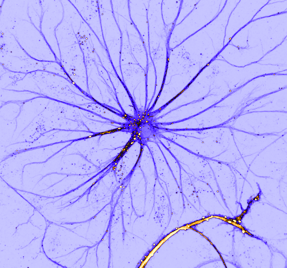
Mysterious increase in the motility of these #lysosomes for this #FluorescenceFriday
@DavidMcGrath_9 did is as a control, but after adding a non-stimuli treatment lyso got crazy, maybe #mechanosignaling triggered by shear stress? IDK, but looks👌🏽!
@focalplane_jcs @cellcommlab
6
53
290
5 May 2022
Some methods sections 😅

1