
Light microscopist, cell biologist. Director @DukeU Light Microscopy Core Facility (LMCF);@Stanford and @AmherstCollege grad; opinions my own.
Joined October 2019
- Tweets 1,174
- Following 875
- Followers 1,056
- Likes 3,284
52 Photos and videos


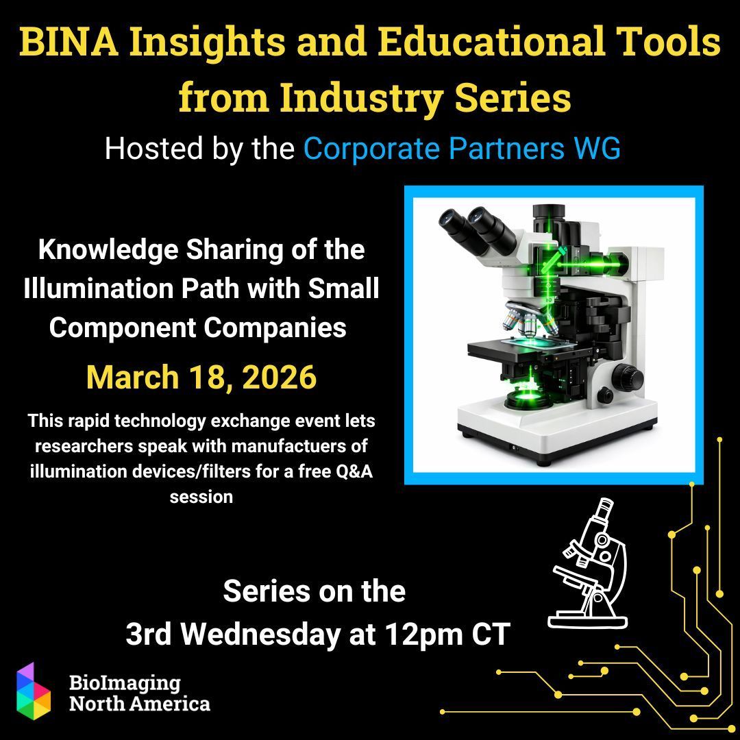
Come learn about, ask questions and give feedback to 10 microscopy component companies: AVROptics-CoolLED-Chroma-89North(a company of Oxxius)-Lumencor-Vortran-TopticaUSA-Coherent-Excelitas-SpectraPhysics ~ March 18 13:00 easternUS ~ bioimagingnorthamerica.org/e…

#HappyFluorescenceFriday #microscopycommunity- join the BINA Corporate Partners for discussions on illuminations devices/filters with manufacturers, March 18. See the website for participating companies
Learn more: buff.ly/UY0TyYE
86
Join us for this educational presentation about fluorescent probes @BioimagingNA bioimagingnorthamerica.org/e…
80
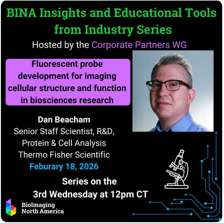
Please join us @BioimagingNA Insights and Educational Tools session Feb 18 at 13:00 easternUS for a virtual talk by Dan Beacham @fishersci Register: bioimagingnorthamerica.org/e…
60
Dr. Lisa Cameron retweeted

#HappyFluorescenceFriday #microscopycommunity - Join the conversation on Fluorescent probe development & best practices in live cell imaging assay development, with Dan Beacham, Senior Staff Scientist at Thermo Fisher Scientific, Feb 18.
Learn more: buff.ly/glYvpuj

2
3
292

21 Oct 2025
Support a scientist for congress in North Carolina - NC11
20 Oct 2025

A scientist from UNC Chapel Hill is running for congress in NC for a seat currently held by a republican. If you care about science (and democracy) please consider donating to his campaign
secure.actblue.com/donate/pa…
1
141
Dr. Lisa Cameron retweeted

20 Oct 2025
A scientist from UNC Chapel Hill is running for congress in NC for a seat currently held by a republican. If you care about science (and democracy) please consider donating to his campaign
secure.actblue.com/donate/pa…
5
14
1,173
Dr. Lisa Cameron retweeted

21 Jan 2025
Applications for our Quantitative Imaging From Acquisition: to Analysis 2025 course @QIatCSHL (Mar 24-Apr 8) are due on Jan 31! . Come immerse yourself in microscopy and image analysis with me, @TalleyJLambert, @florianjug, @BoSoxBioBeth & Hunter Elliott! Please RT! @cshlcourses
14
22
1,995
15 Nov 2024
See you at the Cerulean sky; look for @cameronscope.bsky.social
2
121
18 Sep 2024
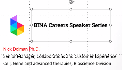
Join @BioimagingNA NOW to hear more about Nick Dolman's career - go to BINA website bioimagingnorthamerica.org/e…
1
184
7 Sep 2024
Congrats to @alsina_fc and @TheSilverLab for nice work and imaging. And thank you from the @DukeU Light Microscopy Core Facility for the acknowledgement.
7 Sep 2024

I am thrilled to share our new publication!! Using mice, organoids, live imaging and biochemistry, we show the RNA binding protein EIF4A3 directly binds and controls microtubules to promote axon growth. Thanks to super talented @alsina_fc and an outstanding team of collaborators!
2
1
7
409
18 Aug 2024
Congrats Chengcheng. We in the Duke LMCF (LightMicroscopyCoreFacility) are glad to continue to help with microscopy imaging & appreciate the conscientious LMCF use by the @NiccoloTerrando lab.
17 Aug 2024

Congrats Chengcheng Song, postdoc in @NiccoloTerrando lab @Duke_Anesthesia @DukeMedSchool for receiving the Alzheimer’s Association Research Fellowship (AARF) to study the impact of #delirium on AD. Impromptu celebration and proud mentor 👏 🎉 @alzassociation @ISTAART #ENDALZ

1
1
7
594

3 Jul 2024
2 Jul 2024

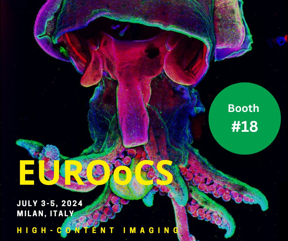
Come to our booth at #EUROoCS in #Milan, Italy, tomorrow!
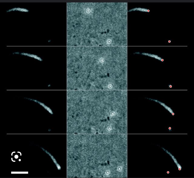
Image Credits: Hairy Squid by M. Estermann, D. Bressan de Andrade, M. Flores Flores, L. de Oliveira Saad. 3D projection of an embryo of Doryteuthis pealeii squid. #CSU #Microscopy Technology in a W1 #Olympus microscope.
2
12
1,027
Dr. Lisa Cameron retweeted

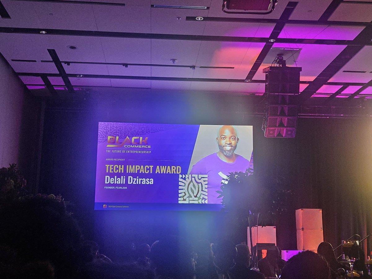
22 Jun 2024
Congrats to the kid. He stays collecting the awards. 💪🏿💪🏿💪🏿

2
5
43
4,988
8 Jun 2024
Yes! thanks so much for all the support! @LeicaMicro especially Louise Bertrand @zeiss_micro and Chris Bjornsson @Yokogawa and @NikonUSA @HamamatsuPhoton @AndorTechnology and some lightsheet systems from @LifeCanvasTech @bruker Thx SUTTER Inst and @Okolabgroup
5 Jun 2024

A big shout out to all the vendors who lend their cutting edge microscopes and equipment to the @MBLScience and #embryo24!
@zeiss_micro @LeicaCameraUSA @NikonUSA @HamamatsuPhoton @Yokogawa @LifeCanvasTech @AndorTechnology @MarketingSutter @Okolabgroup @bruker @Tokai_Hit

12
511
7 Jun 2024
Great to have James embedded in the #embryo24 @MBLScience course. And thanks to @Yokogawa for arranging with the following companies to set up a spinning disk confocal for us @NikonUSA @vortranlaser @HamamatsuPhoton @Okolabgroup @Excelitas
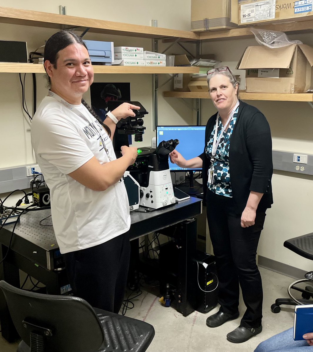
4 Jun 2024

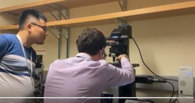
.@SocDevBio Choose Development Fellow James Russette @montanastate training on confocal w/Lisa Cameron @DukeU at Embryology course @MBLScience
1
11
2,216
Dr. Lisa Cameron retweeted

4 Jun 2024
.@SocDevBio Choose Development Fellow James Russette @montanastate training on confocal w/Lisa Cameron @DukeU at Embryology course @MBLScience
1
28
3,774
27 May 2024
Thanks to @WorkingatDuke for highlighting our assistance to @DukeU @DukeMedSchool researchers for their light microscopy needs in the @DukeU Light Microscopy Core Facility. Let us know if we can assist you microscopy.duke.edu We have equipment in FFSC, LSRC, NanDuke&MSRB-III
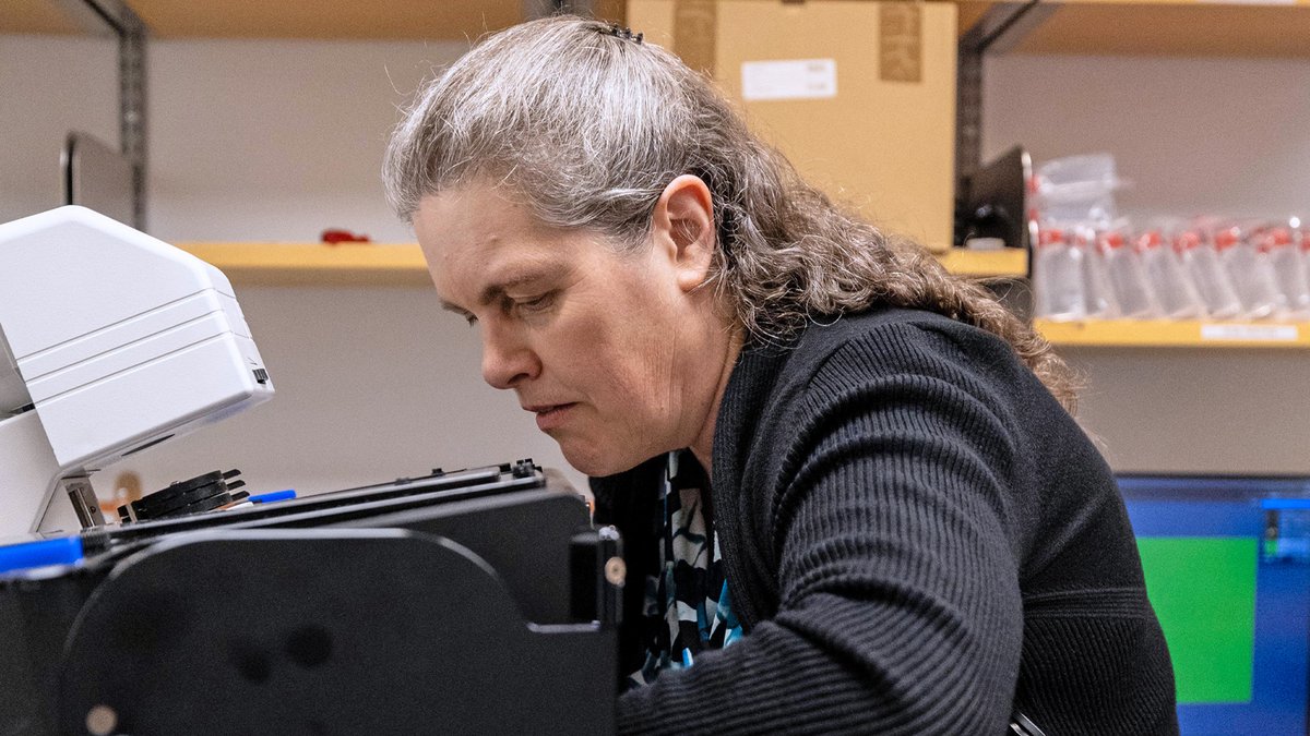
21 May 2024

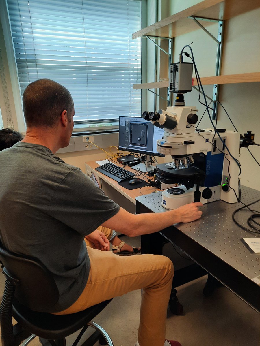
Lisa Cameron is the Director of Duke’s Light Microscopy Core Facility that’s open 24 hours a day, seven days a week in Medical Science Research Building III for researchers to use high-tech microscopes. Read more about her work: stories.duke.edu/a-24-hour-w…
7
36
3,305
Dr. Lisa Cameron retweeted

23 Apr 2024
Excited to be a part of this webinar with renowned experts Jeremy Burrows (MMV) and Lyn-Marié Birkholtz (U of Pretoria, South Africa) on World Malaria Day! -ERD 🌎🦟 acs.org/acs-webinars/library…
(Thursday, April 25, 2024 @ 11am-12:30pm ET. Free to register with ACS ID)
1
5
786
Dr. Lisa Cameron retweeted
23 Apr 2024
We've got another 4-pack of PNC Triangle Club tickets to give away for Thursday night's game thanks to @PNCBank!
Repost for your chance to win!
(winner chosen at noon ET tomorrow, 4/24)
ALT PNC Triangle Club Giveaway graphic.
2
199
77
12,729