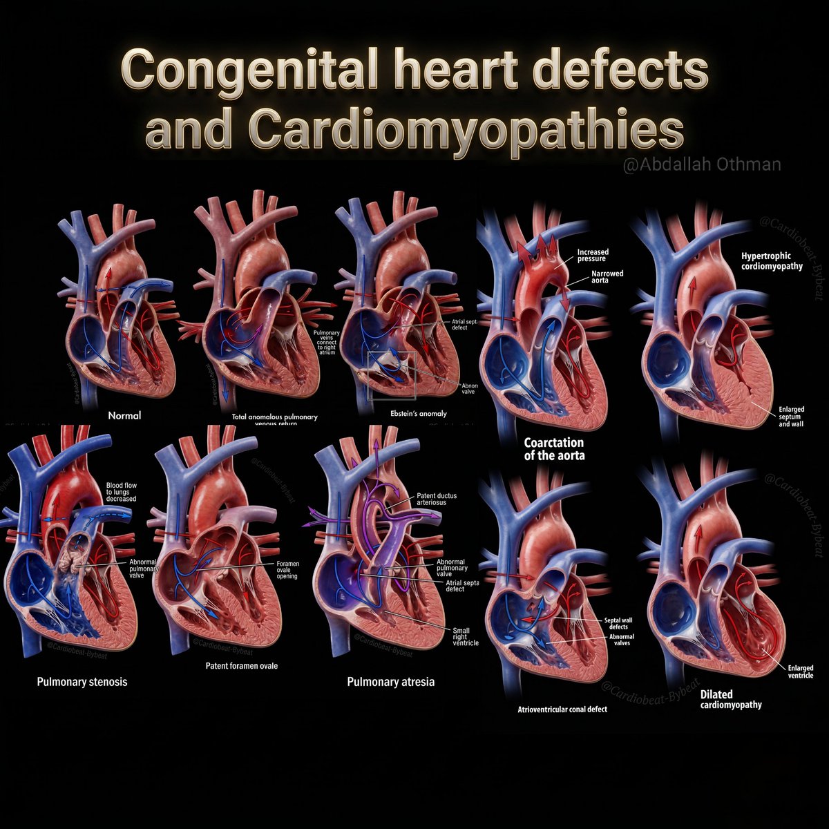
ALT "High-resolution 3D medical illustration comparing 10 major congenital heart defects and cardiomyopathies on a black background. Includes Normal heart anatomy, Total anomalous pulmonary venous return, Ebstein’s anomaly, Coarctation of the aorta, Hypertrophic cardiomyopathy, Pulmonary stenosis, Patent foramen ovale, Pulmonary atresia, Atrioventricular canal defect, and Dilated cardiomyopathy. Detailed labels show key anatomical abnormalities, blood flow changes, and structural defects. Educational infographic for cardiology and medical education
Every day, around 12 people in Northern Ireland die from cardiovascular disease, with the condition affecting more than 230,000 people here.
Our #redbenchdiaries visited Stormont, where @deborah_cheryl and @mcguigan_philip shared the impact of living with cardiovascular disease.
💕 ISUOG's Every Little Heart Matters campaign isn't just about raising awareness. It is also about measuring impact. It is crucial to see the real life impact of the work that we are doing to educate clinicians on the skills needed to accurately detect fetal cardiac anomalies through prenatal ultrasound.
👩⚕️ We spoke to doctors in Oman on the impact of ISUOG outreach, specifically for enhancing their skills in detecting of fetal cardiac anomalies. Here is what Dr Ahlam Al Kalbani, an obstetrician and head of the ultrasound unit at Royal Hospital, Muscat, Oman, had to say:
🗣️ "Because of the ISUOG outreach, we don't just scan with our hands anymore. We scan with a standard of global excellence. It has truly enhanced our ability to protect the smallest hearts in our care."
Find out more about #EveryLittleHeartMatters on the ISUOG website: bit.ly/42jbYL9#CHDAwarenessMonth#CHD#CHDAwareness#HeartMonth#PregnancyCare#LoveUltrasound#Pregnancy#Birth#HeartDefects#HeartHealth#PrenatalDiagnostics#OBGYN#Ultrasound#Obstetrics#Obstetrician
Using human pluripotent stem cell and mouse models of Down syndrome, researchers at @UCSF identified HMGN1, a nucleosome-binding epigenetic regulator encoded on chromosome 21, as a key contributor to the cardiac developmental abnormalities associated with this aneuploidy.
#Trisomy21#HeartDefects#MedicalResearchnature.com/articles/s41586-0…
📓 From the #THIJournal
Angelini, et al. review coronary myocardial bridging, including the diagnostic testing and treatment approaches for symptomatic patients with this most common congenital cardiac anomaly: doi.org/10.14503/THIJ-23-830…#THIPubs#heartdefects
🫀Coarctation of the aorta (CoA)is a potentially life threatening congenital heart defect where unfortunately 60% are not detected before birth.
🩺This group have used AI to assess the baby's heart at the routine 18-22 week scan and have shown significant improvement in detection of CoA.
This is how AI can be introduced into medical practice - being able to automate measurements and identify at risk groups for management.
##MedAI#FetalMedicine#Ultrasound#AIinHealthcare#heartdefects#CHD
Role of artificial‐intelligence‐assisted automated cardiac biometrics in prenatal screening for coarctation of aorta - Taksøe‐Vester - 2024 - Ultrasound in Obstetrics & Gynecology - Wiley Online Library obgyn.onlinelibrary.wiley.co…
Congenital heart disease, present from birth, refers to a range of heart defects affecting normal function. While the exact cause remains unclear, certain risk factors like maternal infections, smoking, alcohol use, and poorly controlled diabetes increase the likelihood of this condition. Although we can’t eliminate all risks, following guidelines such as vaccination, folic acid intake, and avoiding harmful substances can reduce the chances of congenital heart disease and ensure a healthier pregnancy.
#CongenitalHeartDisease#HeartHealth#HealthyPregnancy#FolicAcid#RiskFactors#MaternalHealth#HeartDefects#PreventiveCare#HealthyBaby
(Congenital heart disease, pregnancy, maternal health, risk factors, prevention, folic acid, rubella, smoking, diabetes, heart defects)
Ventricular Septal Defects (VSDs) Explained! 🫀
The ventricular septum is divided into four key regions: inlet, trabecular, outlet, and membranous. A VSD is an abnormal opening in this septum, allowing blood to shunt between the LV & RV.
🔍 Types of VSDs & Their Locations:
📌 Perimembranous (pmVSD): Below the aortic valve, near the membranous septum
📌 Muscular (mVSD): Located in the trabecular septum; can be multiple (Swiss-cheese VSD)
📌 Inlet (iVSD): Near the AV valves, often linked to endocardial cushion defects
📌 Supracristal (scVSD): Just below the pulmonic valve, may lead to aortic regurgitation
How Do We Diagnose VSDs?
Echocardiography plays a key role! Different echo views help identify VSDs:
🔹 Parasternal long-axis → Best for pmVSD & mVSD
🔹 Parasternal short-axis → Locates pmVSD (10 o’clock) & scVSD (1 o’clock)
🔹 Apical 4-chamber → Ideal for detecting inlet & apical muscular VSDs
Why does this matter?
VSD size, location, and associated defects determine hemodynamic impact & management! Some close spontaneously, while others require surgical or percutaneous closure.
Understanding VSDs = Better diagnosis & treatment!
#Cardiology#VSD#ECHOFacts#HeartDefects#MedTwitter
ALT A detailed schematic of the ventricular septum and various types of ventricular septal defects (VSDs).
(A) The ventricular septum is divided into four regions: inlet, trabecular, outlet, and membranous.
(B) Different types of VSDs are shown, including perimembranous, muscular, inlet, and supracristal VSDs.
(C) Parasternal long-axis echocardiographic view illustrating a perimembranous VSD (pmVSD) below the aortic valve and a mid-muscular VSD (mVSD).
(D) Parasternal short-axis view at the aortic valve level showing a pmVSD (10 o’clock position) near the tricuspid valve and a supracristal VSD (scVSD) at 1 o’clock near the pulmonic valve.
(E) Parasternal short-axis view demonstrating a muscular VSD.
(F) Apical four-chamber view highlighting an inlet VSD (iVSD) and multiple muscular VSDs (mVSDs). A primum-type atrial septal defect (ASD) is also indicated.
Some #congenital#heartdefects were, at one time, a fatal childhood diagnosis, but better early treatments have transformed it into a condition that needs to be monitored and managed into adulthood.
Read more here: brnw.ch/21wQKXj#AmericanHeartMonth
Samantha discovered her baby's multiple #heartdefects at her 20-week scan. Jaxon was born underwent his first open-heart surgery at just four days old. He is currently preparing for his next surgery - we wish him all the best.❤ Read their story here: ow.ly/3j6W50TyNxK ❤