
Random lab prettiness
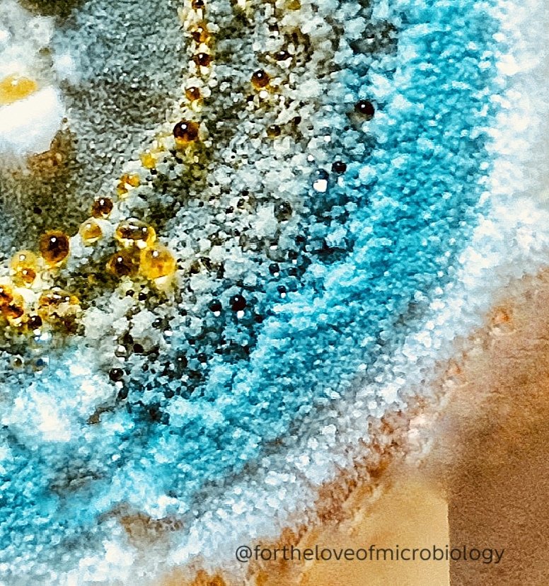
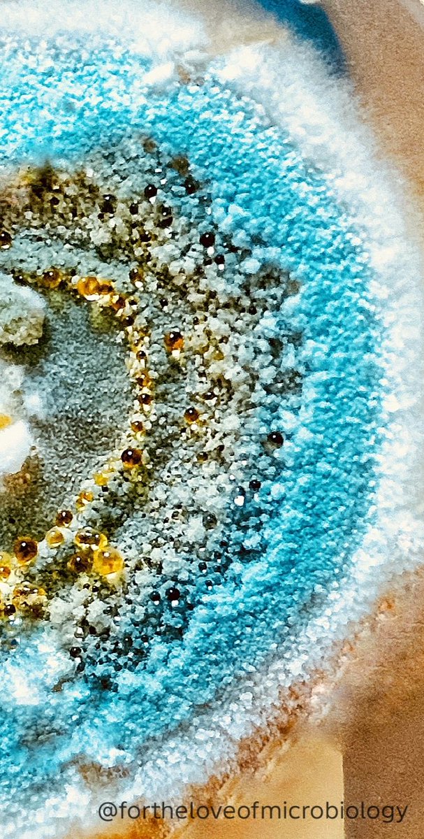
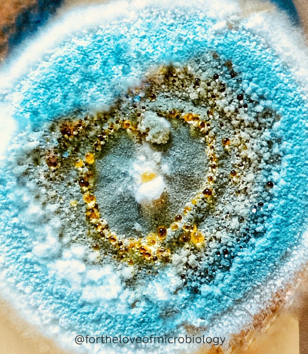
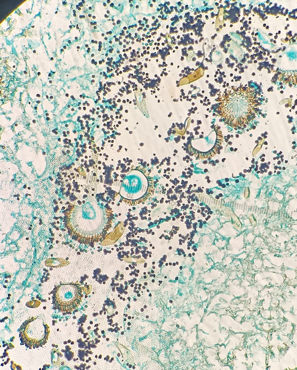
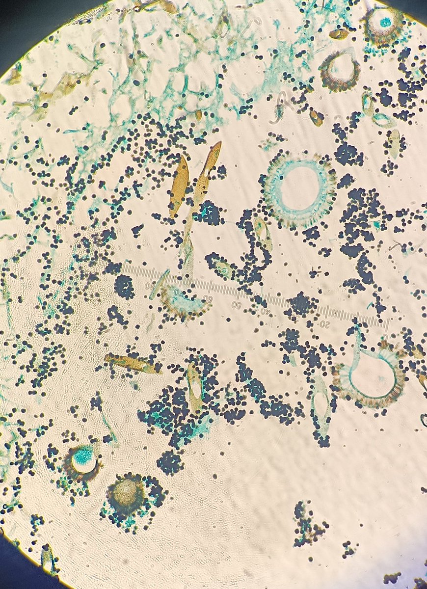
A mature Penicillium colony displaying vibrant blue-green conidiation and abundant honey-colored exudate droplets—a striking example of fungal colony morphology.
#Fortheloveofmicrobiology #clinicalmicrobiology #microrounds #IDpath #ASMClinMicro #STEM #WomeninMicrobiology #ClinMicro #microbiologypakistan #PathBugs
1
5
159
Gabriela retweeted

3
17
57
3,849

Jun 1
May 31

3
243


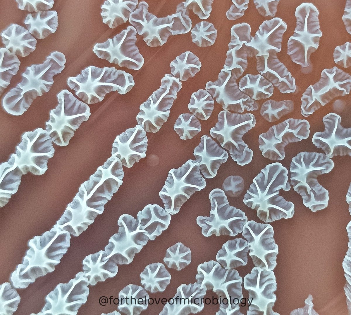
Bacillus subtilis on chocolate agar showing beautifully characteristic large, spreading colonies with a dry, dull, wrinkled colonies. The colony surface displays prominent folds with irregular margins.
This striking colonial morphology is typical of environmental Bacillus species and is often accompanied microscopically by large Gram-positive rods with central to subterminal spores.
B. subtilis is an aerobic, spore-forming bacillus widely distributed in soil, dust, water, and hospital environments. Its spores are highly resilient and can survive under harsh environmental conditions.
In clinical microbiology laboratories, Bacillus subtilis is most commonly encountered as an environmental contaminant or colonizer. However, isolation from sterile sites should not always be dismissed—particularly in immunocompromised patients, patients with indwelling devices, intravenous catheters, prosthetic material, or in the setting of repeated isolation from multiple cultures. Rarely, it has been associated with bacteremia, endocarditis, wound infections, and device-related infections.
One of the fascinating features of B. subtilis is its ability to form complex multicellular communities and biofilms, giving rise to the dramatic wrinkled colony morphology seen here. These surface folds improve nutrient distribution and oxygen exposure within the colony, reflecting the remarkable adaptability of this organism.
#Fortheloveofmicrobiology #clinicalmicrobiology #microrounds #IDpath #ASMClinMicro #STEM #WomeninMicrobiology #ClinMicro #microbiologypakistan #PathBugs
1
7
35
2,350
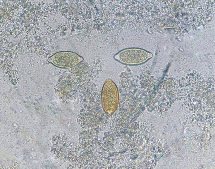
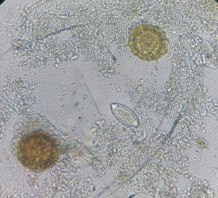
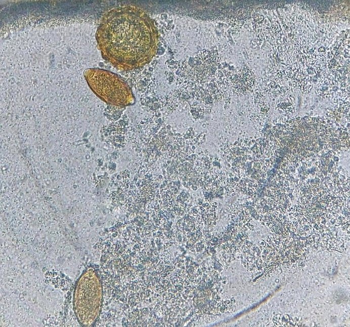
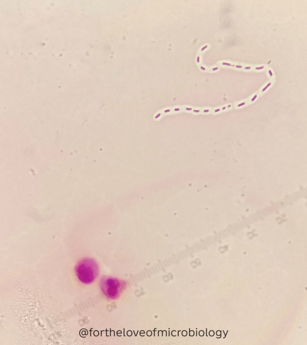
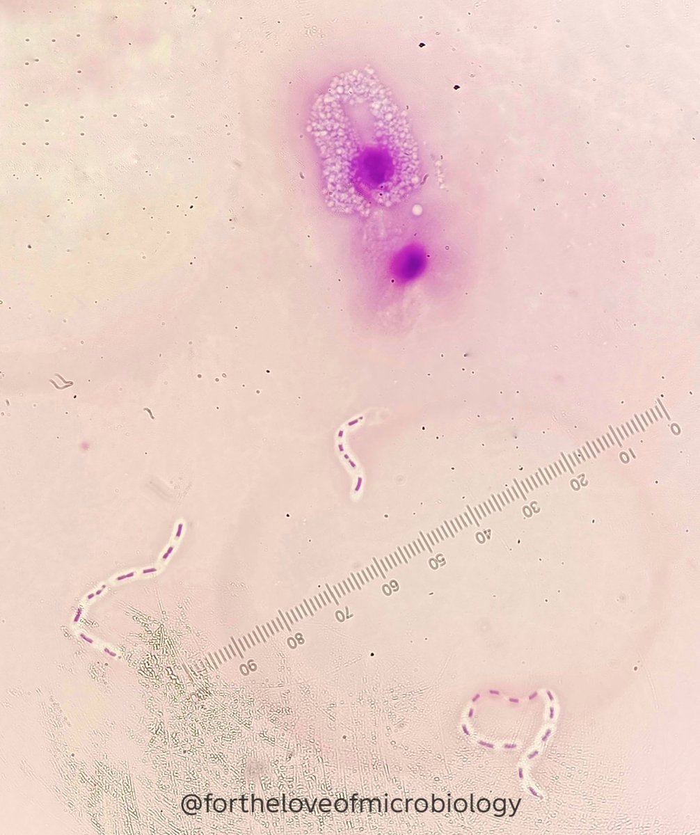
Stool microscopy from a 4-year-old child in Pakistan shows eggs of Ascaris lumbricoides and typical whipworm eggs of Trichuris trichiura in the same sample, confirming a dual parasitic infection.
• Ascaris lumbricoides eggs: Oval with a thick shell and characteristic outer mammillated (corticated) layer
• Trichuris trichiura eggs: Barrel-shaped with characteristic bipolar plugs and a smooth yellow-brown shell
In Pakistan, soil-transmitted helminths are exceedingly common in children—especially where sanitation is poor and access to routine deworming is limited.
• Trichuris trichiura infection may present with mucoid diarrhea, abdominal pain, and iron-deficiency anemia
• Ascaris lumbricoides infection may cause abdominal discomfort, malnutrition, growth impairment, and in heavy infections, intestinal obstruction
Key risk factors in Pakistani children:
• Poverty, rural/peri-urban slum residence, and open defecation
• Unsafe/untreated drinking water and poor sewage disposal
• Inadequate hand hygiene and low caregiver awareness
When a young child presents with chronic anemia, growth faltering, or recurrent gastrointestinal symptoms, stool microscopy for intestinal parasites—including helminth eggs—should be routine. Seeing Ascaris lumbricoides and Trichuris trichiura eggs in the same sample is unfortunately not uncommon in Pakistan and highlights the need for regular deworming, safe water, and strong hygiene education at the family and community levels.
#Fortheloveofmicrobiology #clinicalmicrobiology #microrounds #IDpath #ASMClinMicro #STEM #WomeninMicrobiology #ClinMicro #microbiologypakistan #PathBugs
1
2
8
335
Aspergillus niger on nail tissue, highlighted with GMS stain: septate hyphae and it's beautifully formed conidial head sitting right in the keratinized battlefield.
Not your usual “tinea” – this GMS nail section shows A. niger with a classic conidial head and naturally pigmented brown hyphae and phialides, plus black dot‑like conidia (spores) scattered throughout the slide.
GMS (Grocott–Gomori’s methenamine silver) is a special histology stain that highlights fungi in tissue by turning their cell walls black against a pale background, making even scanty hyphae stand out when H&E struggles.
GMS oxidizes polysaccharides in the fungal cell wall to aldehydes, which then reduce silver ions to black metallic silver, so fungal hyphae and yeasts appear black against a pale green background.
Because of its high contrast and sensitivity, it is especially useful when fungi are scanty or hard to see on routine H&E.
Although dermatophytes dominate onychomycosis, non‑dermatophyte moulds like A. niger are increasingly recognized, genuine nail pathogens – not always “lab contaminants” – especially when repeatedly isolated and seen invading the nail plate.
Non‑dermatophyte mould onychomycosis is under‑diagnosed when we rely only on nail appearance and ignore direct microscopy, repeat cultures and proper identification.
Histology shows the story; mycology confirms the culprit.
Always respect what the stain and the culture are trying to tell you before dismissing that black fungus in the nail.
#Fortheloveofmicrobiology #clinicalmicrobiology #microrounds #IDpath #ASMClinMicro #STEM #WomeninMicrobiology #ClinMicro #microbiologypakistan #PathBugs

2
5
15
720
Not all diagnoses wait for culture.
A patient with a shunt presents with signs of infection.
CSF is sent—and the Gram stain immediately tells a story.
Encapsulated Gram-negative bacilli.
Seen in chains.
A striking capsule, hard to miss.
A very interesting Gram stain—one that needed to be shared.
Culture later confirms Klebsiella pneumoniae.
Clinical context: Ventriculoperitoneal shunt infection.
Clinical pearl:
In CNS device-associated infections, the Gram stain isn’t just preliminary—it can be decisive. A visible capsule is more than morphology—it’s a virulence clue, and should immediately sharpen clinical suspicion and guide early management.
A reminder: sometimes, the most important answer is already on the slide.
#Fortheloveofmicrobiology #clinicalmicrobiology #microrounds #IDpath #ASMClinMicro #STEM #WomeninMicrobiology #ClinMicro #microbiologypakistan #PathBugs

2
4
20
1,748

Mar 16

57. PGY1s, learn this one slide and you will be good to go for 4 major fungal infections. GOLD for boards!!
4
609
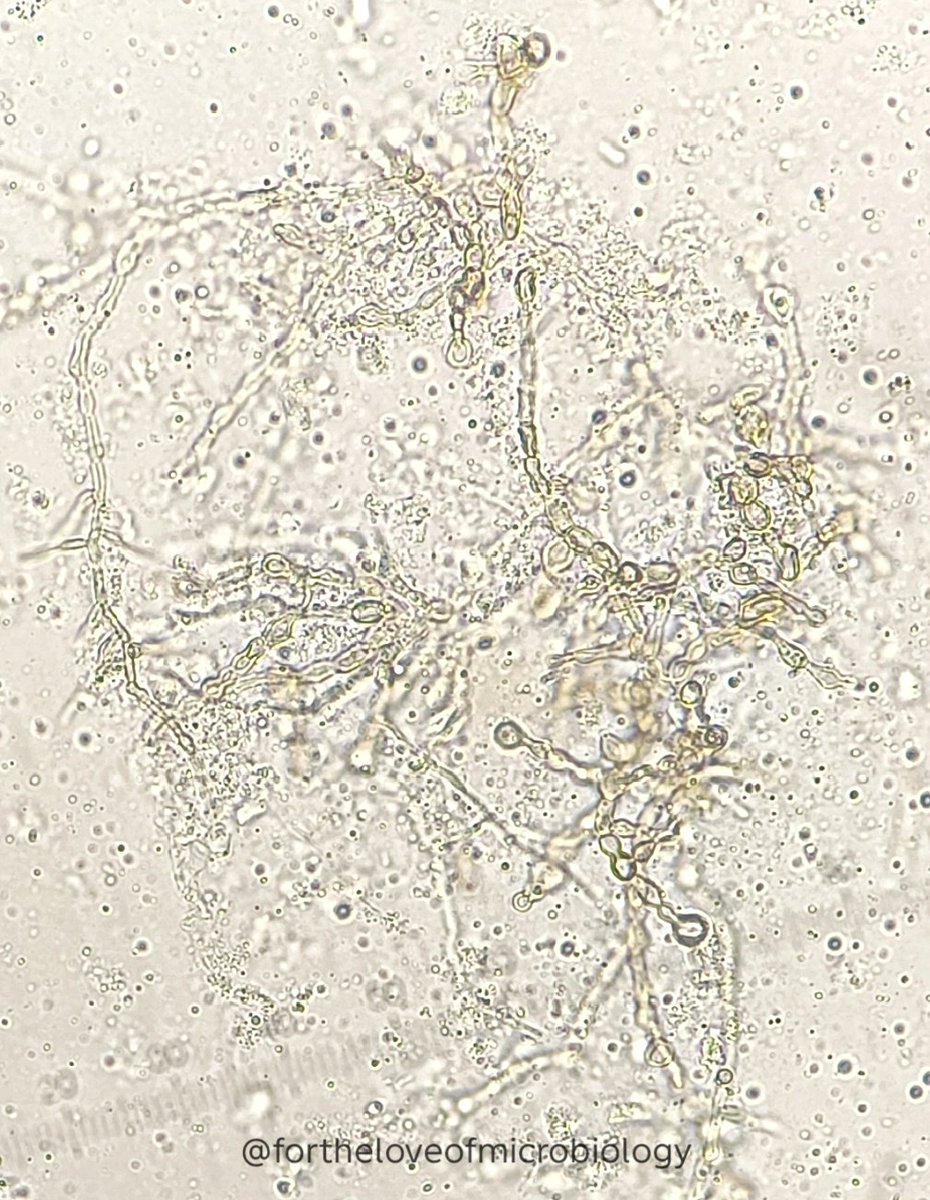
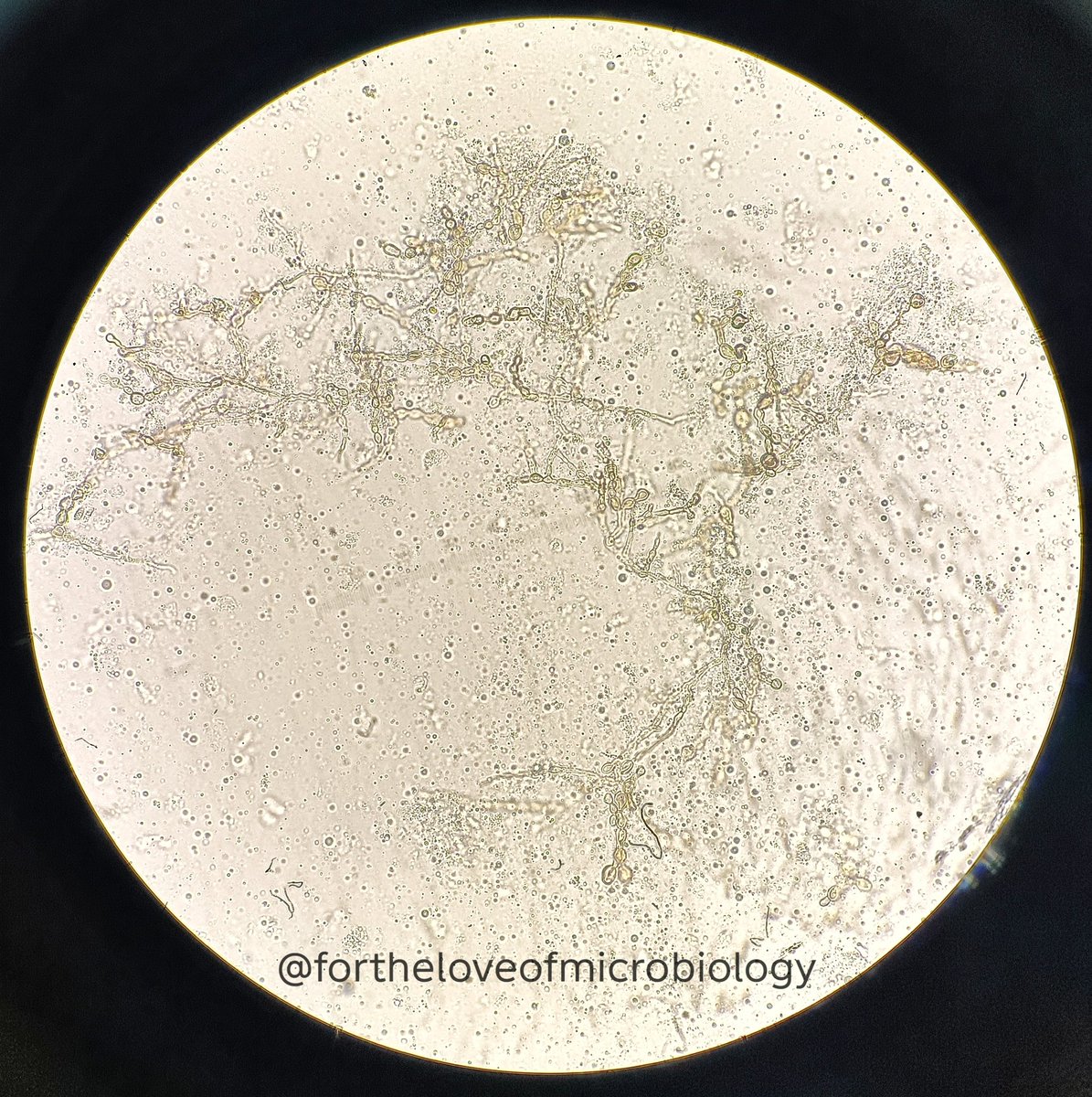
KOH preparation showing pigmented fungal hyphae
When hyphae appear brown on a simple KOH mount, think dematiaceous fungi.
Direct microscopy of the specimen using 10% KOH mount showing septate, pigmented (dematiaceous) fungal hyphae.
The brown pigmentation of the cell wall is due to melanin deposition, a characteristic feature of dematiaceous fungi, which are associated with infections collectively termed phaeohyphomycosis.
Some of the hyphae show a bubble-like or moniliform appearance, reflecting irregular swelling or vacuolation along the hyphal elements. These findings raise suspicion for infection with melanin-producing molds such as Alternaria, Curvularia, Bipolaris, or Exophiala species.
The melanin pigment is not just cosmetic—it is an important virulence factor.
Melanin helps these fungi:
• Protect against oxidative killing by host immune cells (scavenging free radicals)
• Shield against environmental stresses, including UV radiation and enzymatic damage
• Increase resistance to phagocytosis and intracellular killing
This protective effect is one reason why dematiaceous fungi can occasionally cause persistent or invasive infections, particularly in immunocompromised hosts.
Definitive identification requires culture and morphological or molecular confirmation.
#Fortheloveofmicrobiology #clinicalmicrobiology #microrounds #IDpath #ASMClinMicro #STEM #WomeninMicrobiology #ClinMicro #microbiologypakistan #PathBugs
1
2
11
341

Feb 28
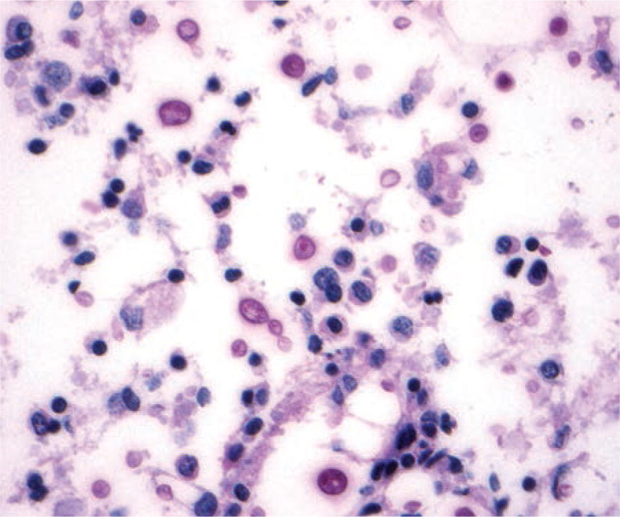
It is not that often that we see these cells/organisms at ROSE (rapid on site evaluation during EBUS)
#pathology #cytopath #idpath #pathbugs
This morphologic finding is more powerful than you think! Please give our latest case report a read if you have a minute. Open access in @DiagnosticCyto 🔥🔥🔥#cytopath
onlinelibrary.wiley.com/doi/…

184
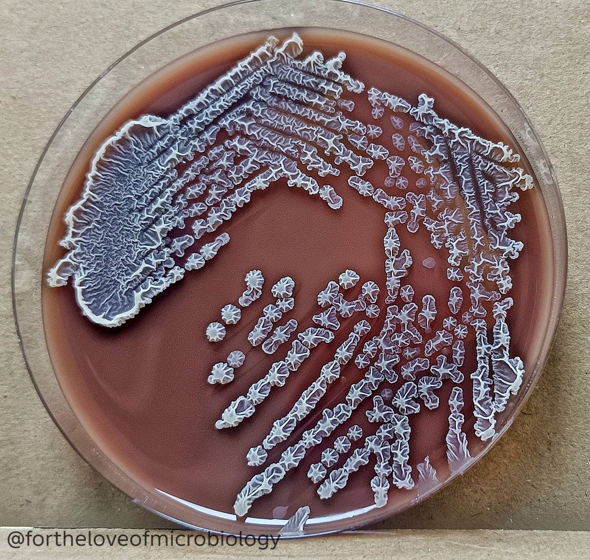
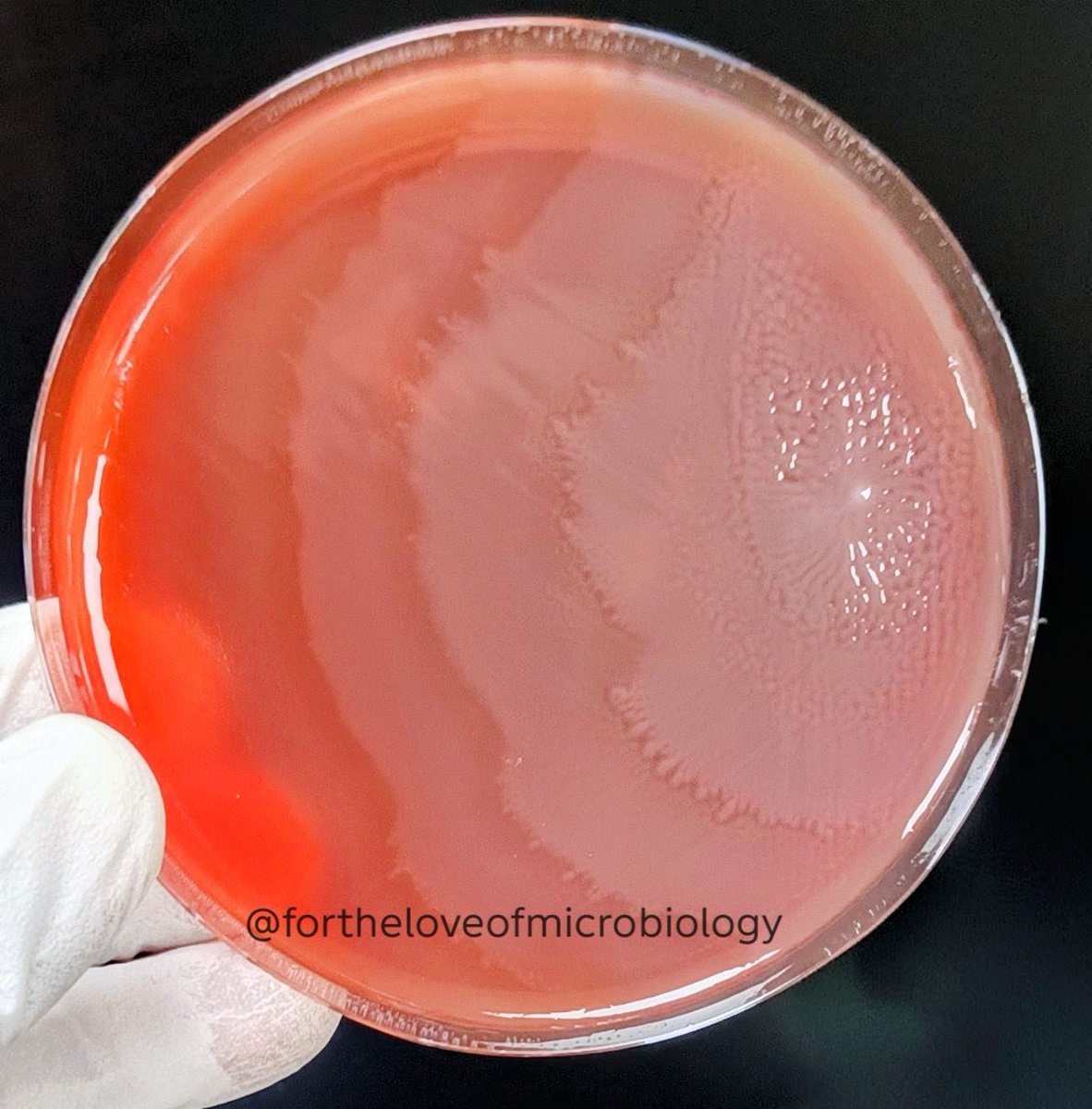
Proteus swarming on a blood agar plate
Proteus is a member of the order Enterobacterales, comprising Gram-negative, facultatively anaerobic bacilli capable of surviving in diverse and nutrient-limited environments. A defining characteristic of Proteus species is their remarkable motility, mediated by numerous peritrichous flagella.
One of the most striking laboratory features of Proteus is the swarming motility, observed as concentric waves of spreading growth across solid agar surfaces, producing the classic “bull’s-eye” or terraced colony pattern on agar plates.
Swarming represents a highly organized, multicellular migration phenomenon driven by cyclic biological differentiation:
🌐 Short vegetative (“swimmer”) cells differentiate into elongated, multinucleate, hyper-flagellated swarmer cells
🌐 Swarmer cells migrate collectively across solid surfaces in coordinated groups or “rafts”.
🌐 Migration is followed by dedifferentiation back into short vegetative cells.
🌐 Cellular multiplication occurs during a resting consolidation phase.
🌐 The cycle then repeats, allowing progressive colony expansion.
Each migration cycle produces a visible growth terrace, resulting in the characteristic concentric ring appearance.
This synchronized population movement represents a form of bacterial social behavior, regulated through environmental sensing and cell-to-cell signaling mechanisms. Surface contact — particularly inhibition of flagellar rotation upon encountering solid media — triggers expression of swarming-associated genes.
The swarmer cell state is not merely a locomotion strategy but is closely linked with virulence:
🌐 Increased production of urease, leading to alkaline urine and struvite stone formation
🌐 Enhanced expression of hemolysin, contributing to tissue damage
🌐 Rapid surface colonization, especially of urinary catheters
Ability to migrate against urine flow, promoting ascending catheter-associated urinary tract infections (CAUTIs)
🌐 Heavy growth may also produce a characteristic fishy or burnt-chocolate odor in culture.
Swarming growth may obscure isolation of other pathogens in polymicrobial specimens.
To suppress swarming and enable colony separation, inhibitory media may be used:
🌐 MacConkey agar
🌐 Deoxycholate citrate agar (DCA) — bile salts inhibit swarming
🌐 CLED agar — electrolyte deficiency prevents swarm differentiation
#Fortheloveofmicrobiology #clinicalmicrobiology #microrounds #IDpath #ASMClinMicro #STEM #WomeninMicrobiology #ClinMicro #microbiologypakistan #PathBugs
3
4
7
541

Feb 22
More from arppress.org/books/book/71:
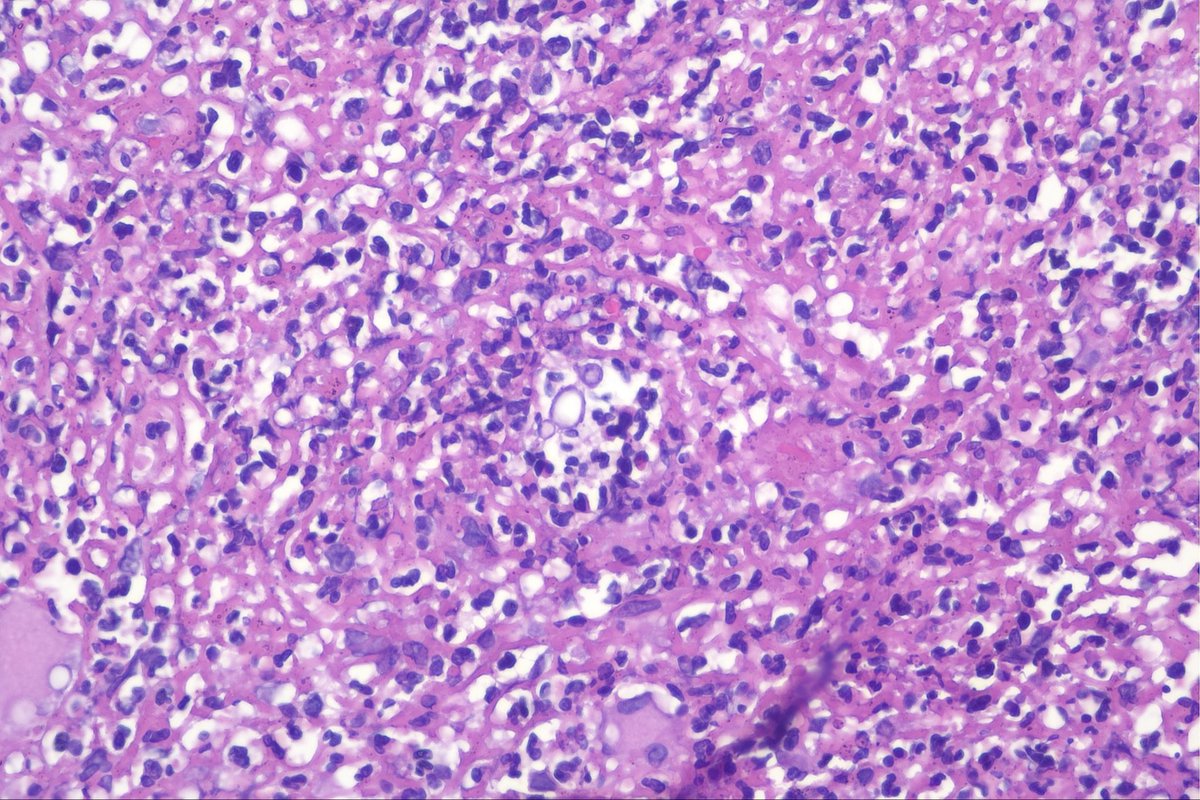
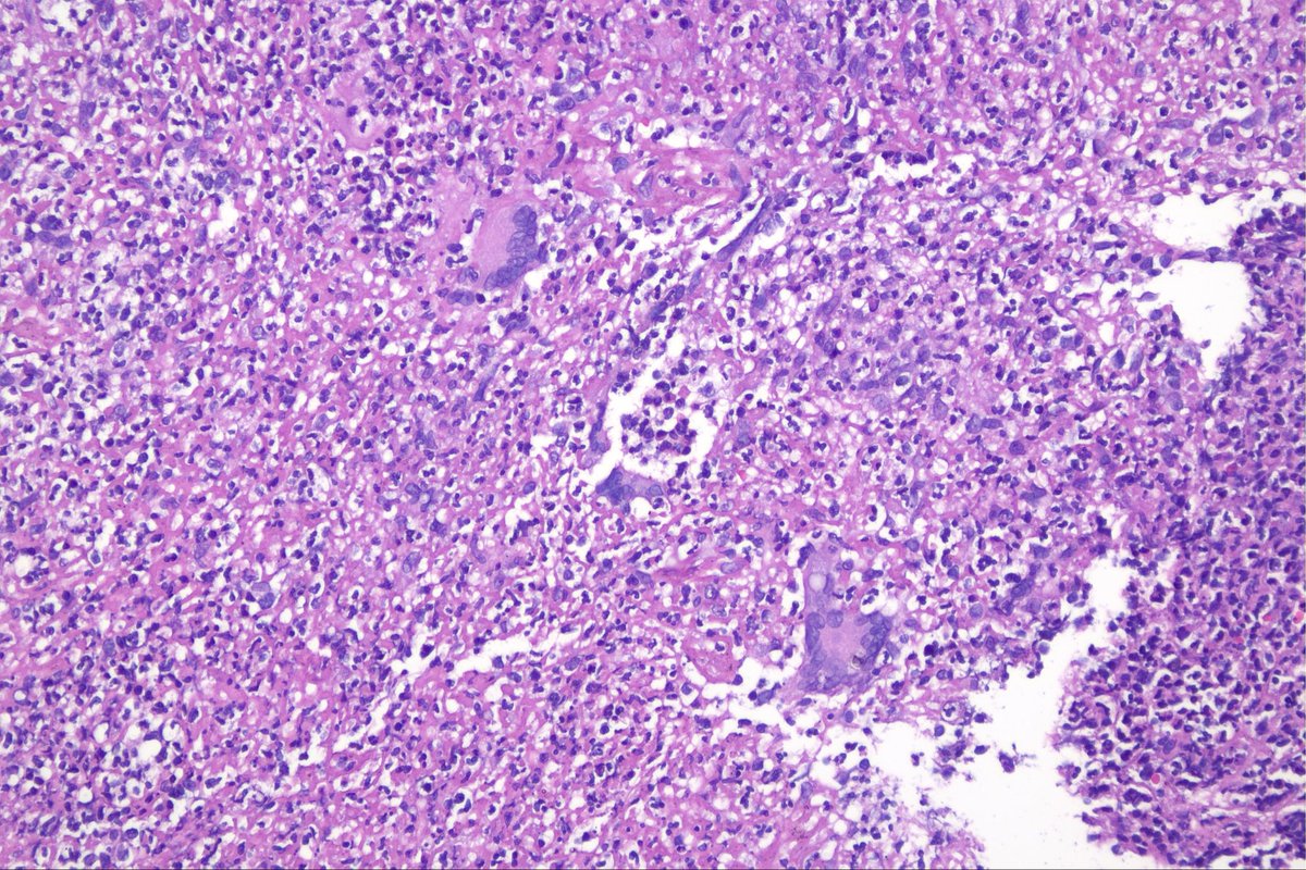
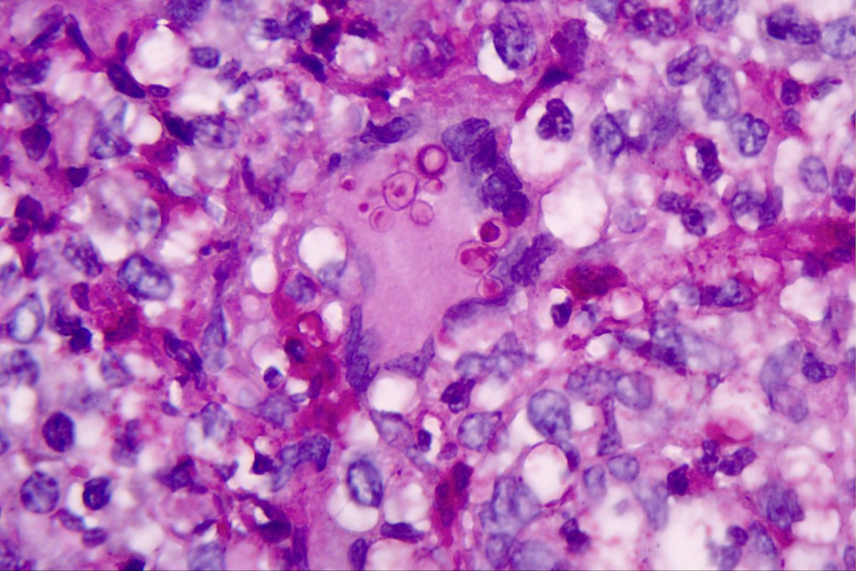
Visceral leishmaniasis (caused by protozoan parasite Leishmania donovani) is endemic to the Mediterranean area, Africa, and South and Central Asia, and usually affects immunosuppressed patients. They are round, 1- to 3-μm organisms found both within histiocytes and extracellularly.
#HemePath #IDpath
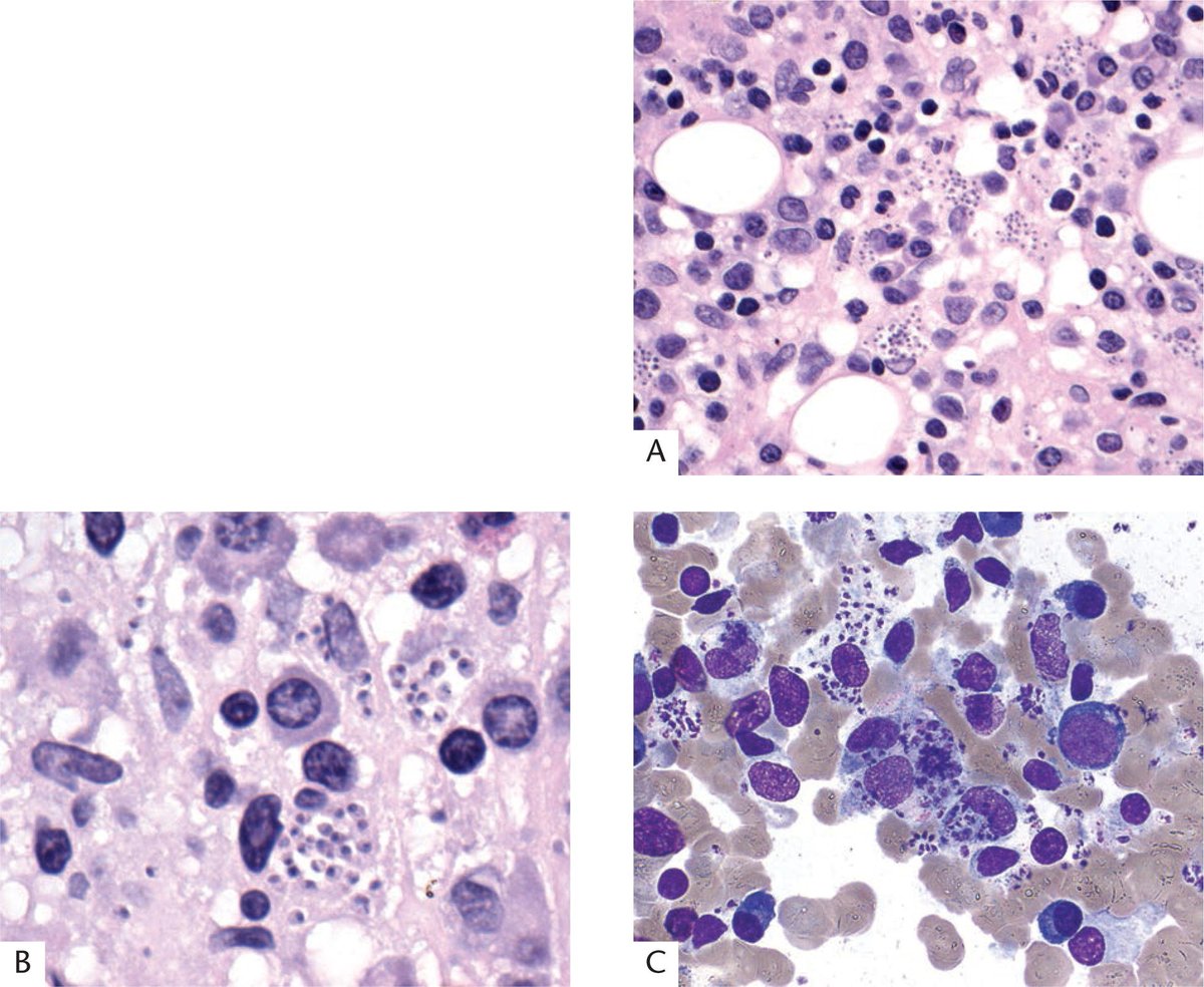
ALT VISCERAL LEISHMANIASIS (KALA AZAR) INVOLVING THE BONE MARROW: In the biopsy specimen, the organisms are intracellular within histiocytes and are often numerous (A,B). The dark nucleus and kinetoplast are visualized in the aspirate smear (C). (Courtesy of Dr. L. de Leval, Lausanne, Switzerland)
2
14
26
1,201

Another cool image from the bone marrow atlas. #pathx #hemepath #pathmatch26 #PathTwitter #idpath @ARP_Press
Feb 18

From arppress.org/books/book/71:
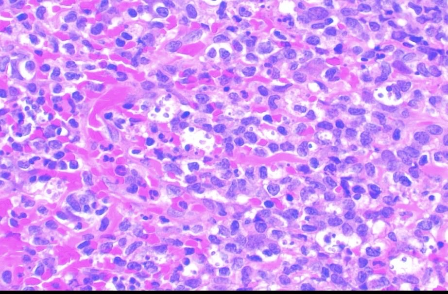
Cryptococcus neoformans involves the bone marrow mainly in the setting of HIV infection, and may be the presenting manifestation with patients often presenting concurrently with meningitis and cytopenias. PAS shown here.
#HemePath #IDpath
ALT CRYPTOCOCCAL INFECTION IN BONE MARROW IN A PATIENT WITH HIV INFECTION: Budding yeast are visible in the bone marrow biopsy. (PAS stain)
1
3
519