
25 Nov 2025
LV Torsion: A Powerful Marker of Systolic Function
Definition
Left ventricular (LV) torsion (or twist) refers to the wringing motion of the heart during systole, apical counterclockwise rotation vs basal clockwise rotation.
It results from the unique helical arrangement of subendocardial and subepicardial fibers.
How is it measured?
Using speckle-tracking echocardiography, torsion is calculated as the net difference in rotation between apex and base, typically in degrees.
We can also measure the rate of rotation (deg/sec), offering further functional insight.
Why does it matter?
Torsion reflects myocardial efficiency and contractile function.
It is sensitive to:
🔵 Early systolic dysfunction (even with preserved EF)
🔵 Myocardial ischemia and infarction
🔵 Cardiomyopathies (HCM, DCM)
🔵 Heart failure (including HFpEF)
Reference:
Modified from Bulwer BE, Solomon SD. In: Atlas of Echocardiography, 2nd ed. Springer; 2009:63.
#Cardiology #Echo #LVFunction #StrainImaging

1
59
181
8,548

28 Jul 2025
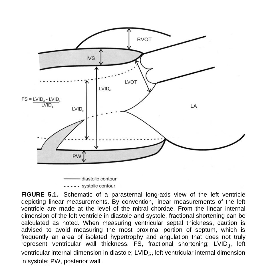
🫀 Hoy revisamos uno de los planos clásicos del #ecocardiograma el eje largo parasternal! La clave para medir bien 🧪 tu ventrículo izquierdo
📐 ¿Qué se mide aquí? 🤷🏽♂️
📏 En el eje largo parasternal, las mediciones #lineales del VI se hacen a nivel de las cuerdas tendinosas mitrales.
🔍 Estas son las estructuras y mediciones principales:
•💆🏽♂️ LVIDd: Diámetro interno del VI en diástole
•💪🏽 LVIDs: Diámetro interno del VI en sístole
•➗ FS: Fracción de acortamiento
FS% = (LVIDd - LVIDs) / LVIDd x 100
•🧱 IVS: Tabique interventricular
•🧱 PW: Pared posterior
•🫁 RVOT: Tracto de salida del ventrículo derecho
•🫀 LVOT: Tracto de salida del VI
⚠️ ¡pero ojo!
📌 No midas la porción más proximal del tabique 🧱, ya que suele haber hipertrofia aislada y angulaciones que no reflejan el grosor real de la pared.
🧙🏼 Este plano es básico para estimar función sistólica, valorar hipertrofias y obtener medidas confiables.🫀
#Ecocardio #POCUS #EjeLargo #UltrasonidoC #EchoTips #EchoBasics #FractionalShortening #LVfunction
1
43
136
7,493

16 Jul 2025
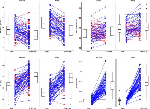
Establishing Cardiac MRI Reference Ranges Stratified by Sex and Age for Cardiovascular Function during Exercise
pubs.rsna.org/doi/epdf/10.11…
#MRI #exercise #LVfunction #SportsCardiology
1
6
826
14 Jun 2025
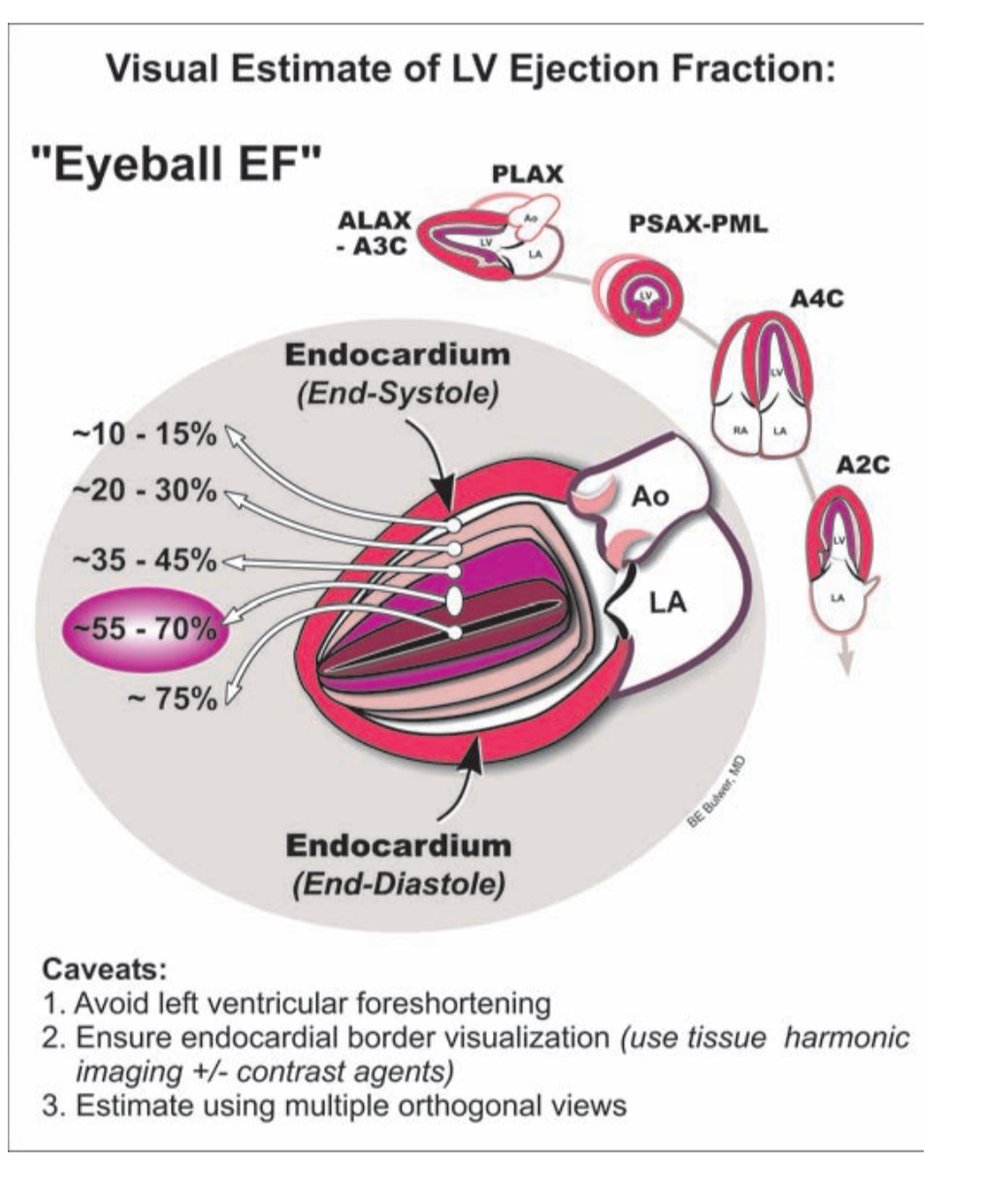
"Eyeball EF" Visual Estimation of Left Ventricular Ejection Fraction
A quick, practical method used in echo labs and at the bedside to estimate LV systolic function using real-time 2D echocardiography.
▶️ Key visual clues: Assess the degree of inward movement and thickening of the LV endocardium during end-systole relative to its size at end-diastole.
🔸 Normal EF (~55–70%):
– Good myocardial thickening
– Vigorous inward motion of walls
– Normal cavity obliteration
🔸 Mildly reduced (~35–45%):
– Wall motion reduced
– Endocardium thickens less
– Cavity smaller but not normal
🔸 Moderately reduced (~20–30%):
– Poor wall thickening
– Sluggish endocardial motion
– LV remains large in systole
🔸 Severely reduced (~10–15%):
– Barely any thickening
– Minimal cavity reduction
– Nearly akinetic walls
🔸 Hyperdynamic (>70–75%):
– Seen in volume depletion or stress states
– Cavity nearly obliterates
Tips for accurate visual estimation:
✅ Avoid LV foreshortening (elongate the chamber fully)
✅ Ensure clear visualization of endocardial borders (use tissue harmonic imaging ± contrast)
✅ Use multiple views: PLAX, A4C, PSAX-PML, A2C, ALAX
Visual EF estimation remains an essential skill for every echocardiographer. Train your eyes and correlate with Simpson’s method when possible!
📖 Ref: Based on concepts from BE Buller, MD (Echo Core Visual Guide)
#Echo #Cardiology #LVFunction #EjectionFraction #MedEd #POCUS #Ultrasound #PointOfCare
5
115
368
43,336
24 May 2025
LV Torsion: A Powerful Marker of Systolic Function
Definition
Left ventricular (LV) torsion (or twist) refers to the wringing motion of the heart during systole, apical counterclockwise rotation vs basal clockwise rotation.
It results from the unique helical arrangement of subendocardial and subepicardial fibers.
How is it measured?
Using speckle-tracking echocardiography, torsion is calculated as the net difference in rotation between apex and base, typically in degrees.
We can also measure the rate of rotation (deg/sec), offering further functional insight.
Why does it matter?
Torsion reflects myocardial efficiency and contractile function.
It is sensitive to:
🔵 Early systolic dysfunction (even with preserved EF)
🔵 Myocardial ischemia and infarction
🔵 Cardiomyopathies (HCM, DCM)
🔵 Heart failure (including HFpEF)
Reference:
Modified from Bulwer BE, Solomon SD. In: Atlas of Echocardiography, 2nd ed. Springer; 2009:63.
#Cardiology #Echo #LVFunction #StrainImaging
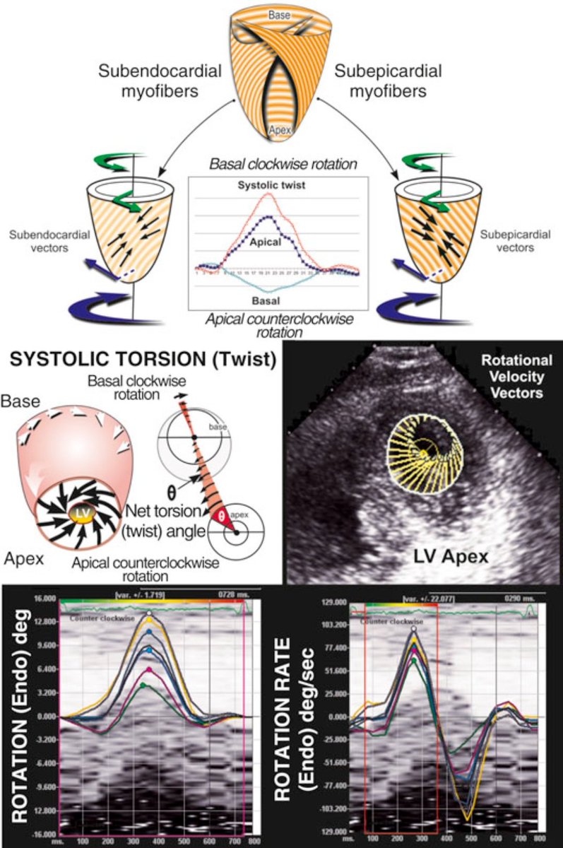
ALT Diagram and echo images illustrating left ventricular (LV) torsion. The top half shows how subendocardial fibers cause apical counterclockwise rotation and subepicardial fibers cause basal clockwise rotation during systole. The center section explains net systolic twist (torsion) as the angle difference between apex and base. The bottom half includes echocardiographic speckle-tracking images and graphs showing LV rotation (in degrees) and rotation rate (degrees/sec) during the cardiac cycle.
1
82
285
23,539
3 May 2025
M-Mode Echo: A Window into LV Function
This schematic illustrates how 2D-guided M-mode echocardiography measures left ventricular (LV) wall thickness and chamber dimensions throughout the cardiac cycle.
Timing of Measurements:
- Diastole: Measured at the Q wave of the ECG
- Systole: At max posterior septal motion, when motion is normal
Key Parameters:
- LVIDd = LV internal diameter in diastole
- LVIDs = LV internal diameter in systole
- STd/STs = Septal thickness (diastole/systole)
- PWTd/PWTs = Posterior wall thickness (diastole/systole)
What is Fractional Shortening (FS)?
Fractional shortening (FS) is the percentage change in LV internal diameter from diastole to systole.
Formula
FS (%) = [(LVIDd − LVIDs) ÷ LVIDd] × 100
Normal range: 25–45%
Clinical Significance:
- Reduced FS% suggests impaired LV systolic function (e.g., in heart failure or cardiomyopathy)
- Increased wall thickness may point to LV hypertrophy (e.g., due to hypertension or aortic stenosis)
- Rapid and reproducible—ideal for screening and serial assessments
Source: Aurigemma GP et al., Curr Probl Cardiol. 1995;20:381.
#Cardiology #EchoFirst #MedEd #CardioTwitter #LVFunction

1
38
243
24,816
2 May 2025
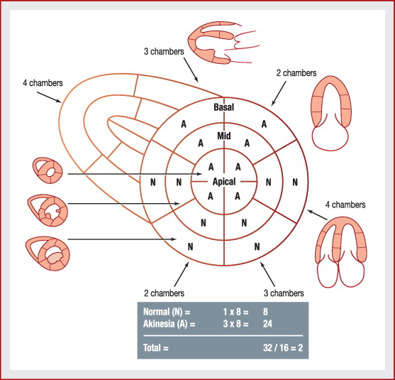
Wall Motion Score Index (WMSI): A key tool in echocardiography
WMSI is used to assess segmental LV wall motion abnormalities often in ischemic heart disease.
Wall segments are graded qualitatively:
1️⃣ Normal: Normal inward motion & thickening
2️⃣ Hypokinesis: Reduced motion (<5 mm), delayed contraction
3️⃣ Akinesis: No inward motion (<2 mm) or thickening
4️⃣ Dyskinesis: Outward/bulging motion (thin, scarred myocardium)
(Some add Aneurysmal: diastolic contour abnormality)
WMSI = Total segment scores / Number of segments visualized
Higher WMSI correlates with more severe LV dysfunction & worse prognosis.
Source: Catherine m otto clinical textbook of echocardiography
#CardioTwitter #Echo #Cardiology #LVFunction #MedEd
1
32
150
8,150
12 Apr 2025
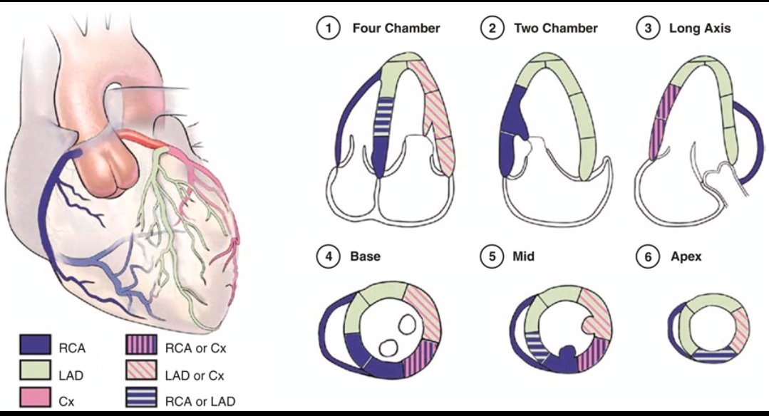
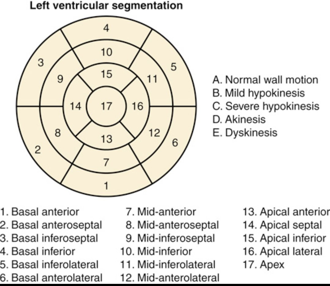
Understanding regional wall motion abnormalities (RWMA) starts with this: the 17-segment LV model.
It divides the left ventricle into basal, mid, apical segments for precise localization of dysfunction.
Why does it matter?
RWMA refers to areas of the LV wall that contract abnormally—key in diagnosing ischemia, infarction, cardiomyopathies, and evaluating viability.
Wall motion grades: A. Normal
B. Mild hypokinesis (↓ motion)
C. Severe hypokinesis
D. Akinesis (no motion)
E. Dyskinesis (paradoxical motion)
Clinical significance:
- Helps identify territory of ischemia/infarct (e.g., LAD vs RCA vs LCx).
- Essential for assessing outcomes post-MI or revascularization.
- Guides decisions in cardiac surgery, ICD implantation, and heart failure management.
@TrackYourHeart
#Cardiology #Echocardiography #MedTwitter #LVFunction #RWMA
2
40
138
12,511

3 Apr 2025
#EHJCVI Phenotyping of left ventricular function in non-ischaemic cardiomyopathy: may unsupervised clustering supersede a parametric evaluation? 🤔
In modern cardiology EF alone is insufficient and inconclusive. #Cardiology #LVFunction
doi.org/10.1093/ehjci/jeaf03…
@EACVIPresident
5
7
676
3 Apr 2025
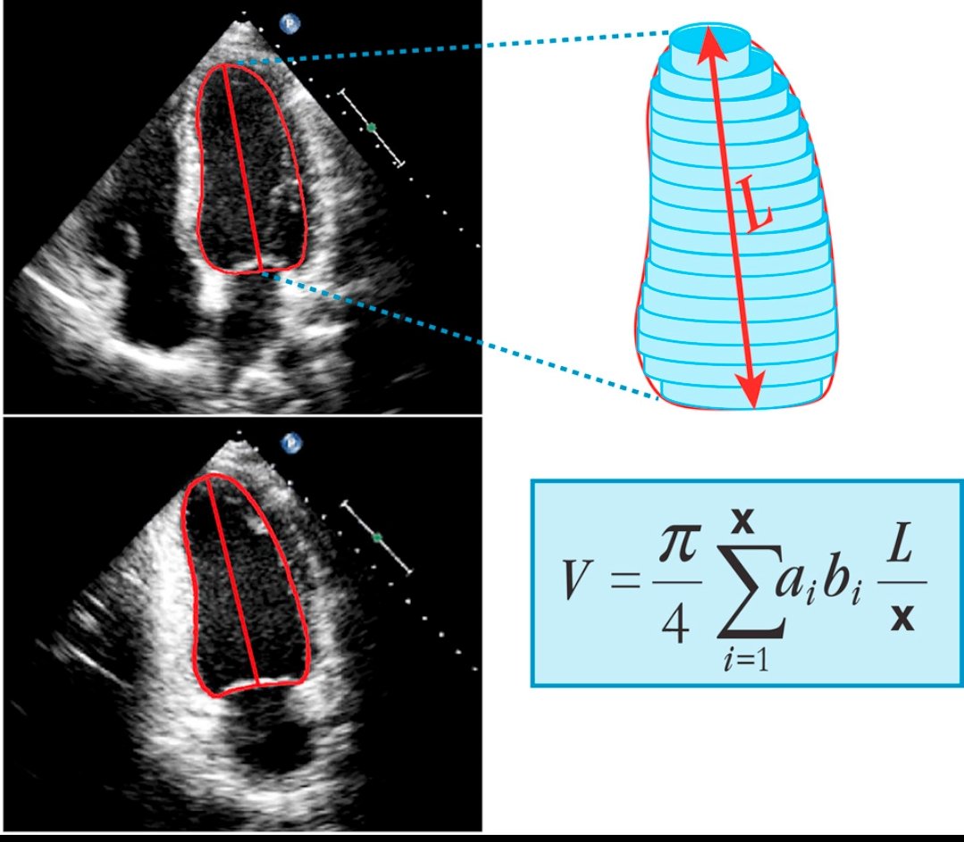
Simpson’s Rule for LV Ejection Fraction Calculation
Ejection Fraction (EF) is the percentage of blood the left ventricle (LV) pumps out with each beat. It is a key measure of cardiac function:
✅ Normal EF: 55–70%
⚠️ Borderline EF: 41–50%
❌ Reduced EF (HFrEF): <40%
Simpson’s Biplane Method is the preferred echocardiographic technique for EF estimation. It calculates LV volume by dividing the ventricle into stacked discs, using:
V = (π/4) * Σ (aᵢ * bᵢ) * (L/x)
- a & b = diameters in apical 4-chamber & 2-chamber views
- L = LV long-axis length
- x = number of discs
Why is EF important?
📌 Low EF suggests heart failure, cardiomyopathy, or ischemic heart disease.
📌 High EF (>75%) may indicate hypertrophic cardiomyopathy.
📌 EF guides treatment decisions, including medications, ICD implantation, and heart failure management.
Accurate EF calculation using Simpson’s method is crucial for diagnosing & monitoring cardiac conditions.
📖 Ref: Lang RM, et al. JASE 2015;28:1-39.
#Cardiology #Echocardiography #HeartFailure #LVFunction
1
68
235
13,859

18 Feb 2025
What's in the left ventricle? 🫀 #echofirst
#EchoFirst #Cardiology #Echocardiography #LVFunction #HeartHealth #POCUS #CardioTwitter #MedicalImaging #LeftVentricle #CardioEcho #FOAMed #HeartFailure #ValvularHeartDisease @GKardiyolog
 Zehra Uyan Ulaş
1
9
37
4,408
28 Oct 2024
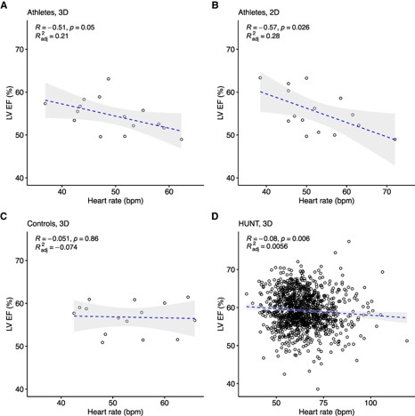
Comparison of resting heart rate and left ventricular ejection fraction in elite endurance athletes and the general population
academic.oup.com/eurjpc/adva…
#Athlete #heartrate #LVfunction #SportsCardiology
4
23
1,158
6 Aug 2024
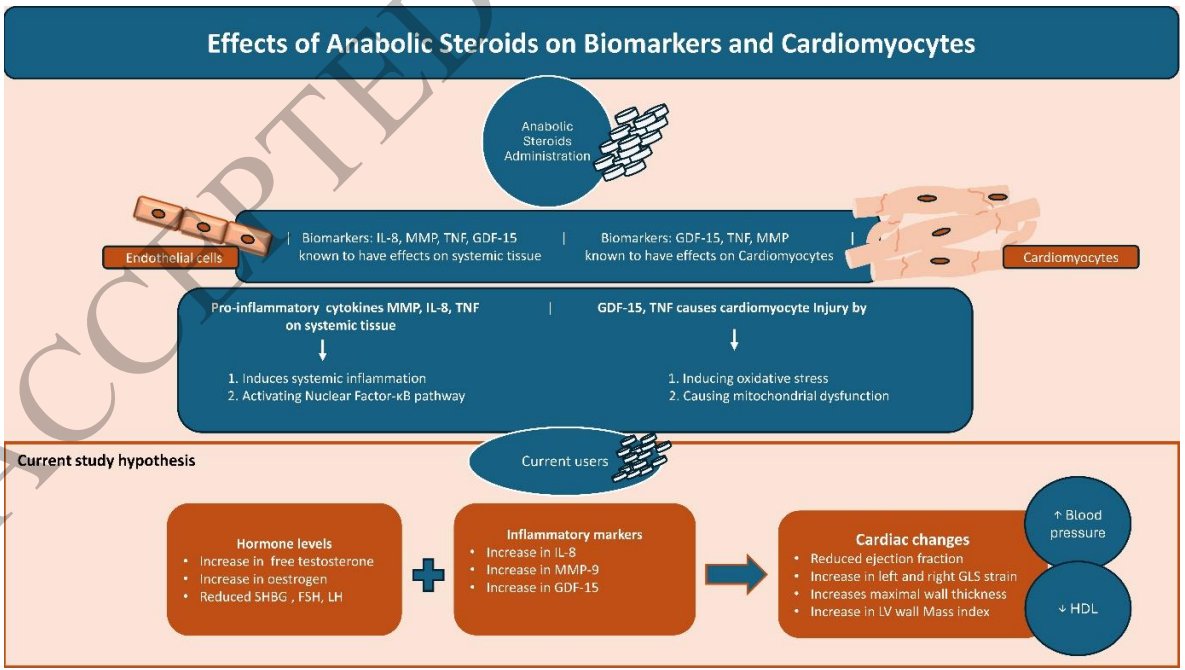
Anabolic steroids in athletes: The interplay of hormones and inflammation leading to the heart's vulnerability
academic.oup.com/eurjpc/adva…
#Steriods #athletes #LVfunction #Sportscardiology
5
9
729

22 May 2024
#Pocus #EPSS #LVFUNCTION #Hemoddynamics #Foamed #FoamCC #ultrasound #Echo
Patients with Systolic Heart Failure (HFrEF) often experience symptoms like shortness of breath, orthopnea, paroxysmal nocturnal dyspnea (PND), exercise intolerance, irregular heartbeats, & edema.
1
3
7
863

14 Apr 2024
Cardiac sarcoidosis:
#1 most asymptomatic. Initial presentation = sudden death!
#2 screen for arrhythmias and LVfunction with ECG, echo, MRI, FDG-PET; repeat ECG yearly
#3 Rx= CS IS > just CS; also cardiac care (GDMT/ EP procedure)
#4 5YS > 90% 10YS> 80%
#SOTA24 @dawn_pedrotty
2
3
8
905

Live today at 12 PM CST for this week's MMI Conference as @maquinonesmd presents “Echo Assessment of Chamber Morphology and LV Function.” #Morphology #Echo #Cardiovascular #MedImaging #Imaging #LVFunction
Watch it Live @ bit.ly/3yz5dWB

1
3
Join us tomorrow at 12 PM CST for this week's MMI Conference as @maquinonesmd presents “Echo Assessment of Chamber Morphology and LV Function.” #Morphology #Echo #Cardiovascular #MedImaging #Imaging #LVFunction
Watch it Live @ bit.ly/3yz5dWB
4

11 Jul 2022
#Cardiotwitter: Robert A. Kloner, MD, et al, have published this article in JACC: Basic to Translational Science on cardioprotective strategies for #STEMI, with a focus on #SSO2 Therapy for reducing infarct size. #MVO #LVfunction sciencedirect.com/science/ar…
1
4

19 Apr 2022
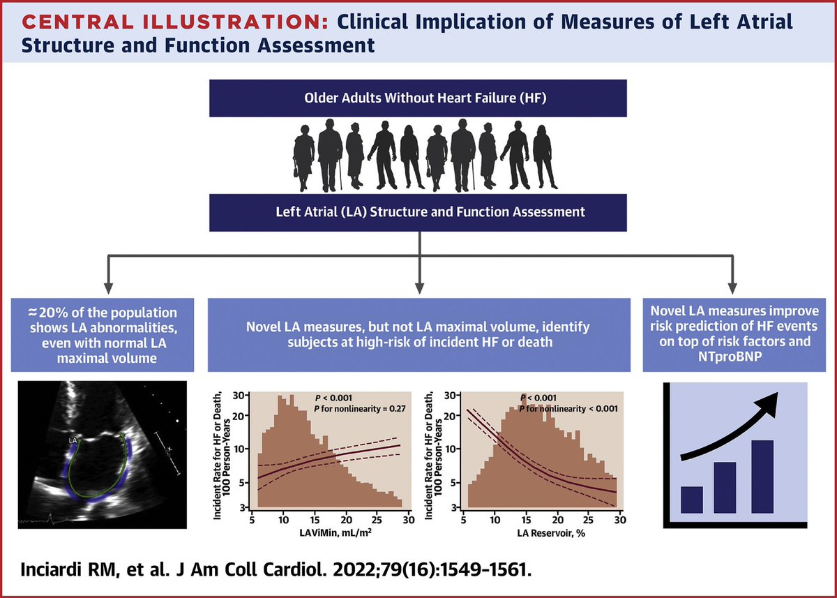
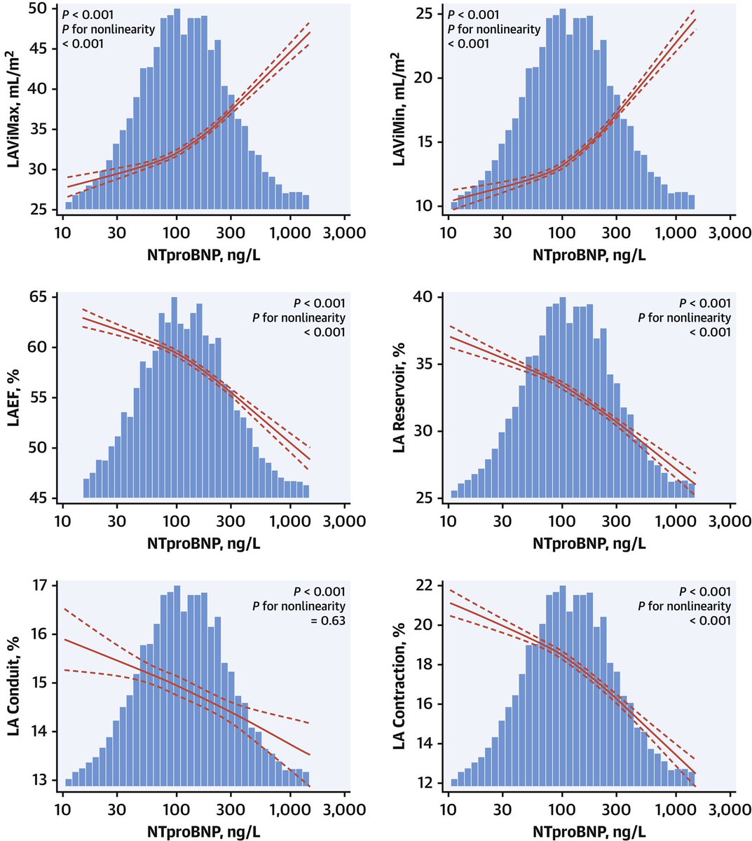
Novel measures of LA structure and function, but not standard assessment by LAViMax, are associated with increased risk of incident #HF or death regardless of measures of LVfunction and #NT-proBNP
#JACC #JACCHF @JACCJournals jacc.org/doi/10.1016/j.jacc.… @scottdsolomon @ShelleyZieroth
21
50

10 Feb 2022
10 Feb 2022

Do you routinely measure #MAPSE?
Did you know that MAPSE beats #GLS and #LVEF regarding prognosis?
A new multicenter collab study out, led by @KellmanPeter
Ref: pubmed.gov/35132872
#WhyCMR 🫀🧲 #EchoFirst 🫀🔊
A thread 🧵. 1/n

3