Jonathan He retweeted

Jun 12
Jun 11

Wondering what your next steps are as a newly matched Resident? Join this member-only roundtable to learn about the support systems available to you and pathways to success. Live instruction, Q&A, and networking opportunities are included; sign up now to give yourself a free career roadmap: bit.ly/42P0Pl6
#ResidentRoundtableSeries #Residency

1
3
324
16 Dec 2025
17 Jul 2025

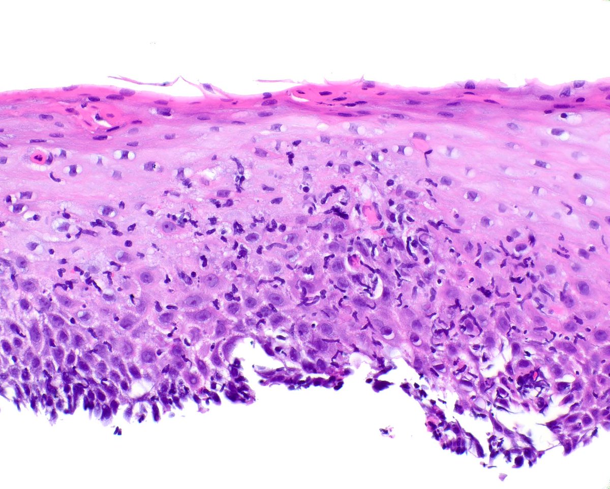
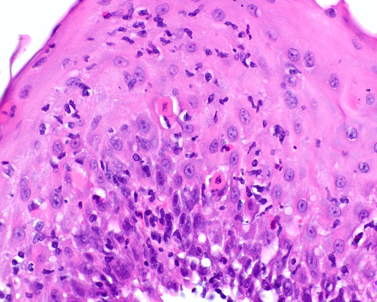
This is an esophagus biopsy with a lymphocyte predominant/lichenoid pattern of injury. Sometimes it correlates with skin lichen planus but more often another type of immunologic alteration is present. This pattern can be accompanied by strictures. Note the squamous apoptosis.
2
13
1,089

19 Nov 2025
18 Nov 2025
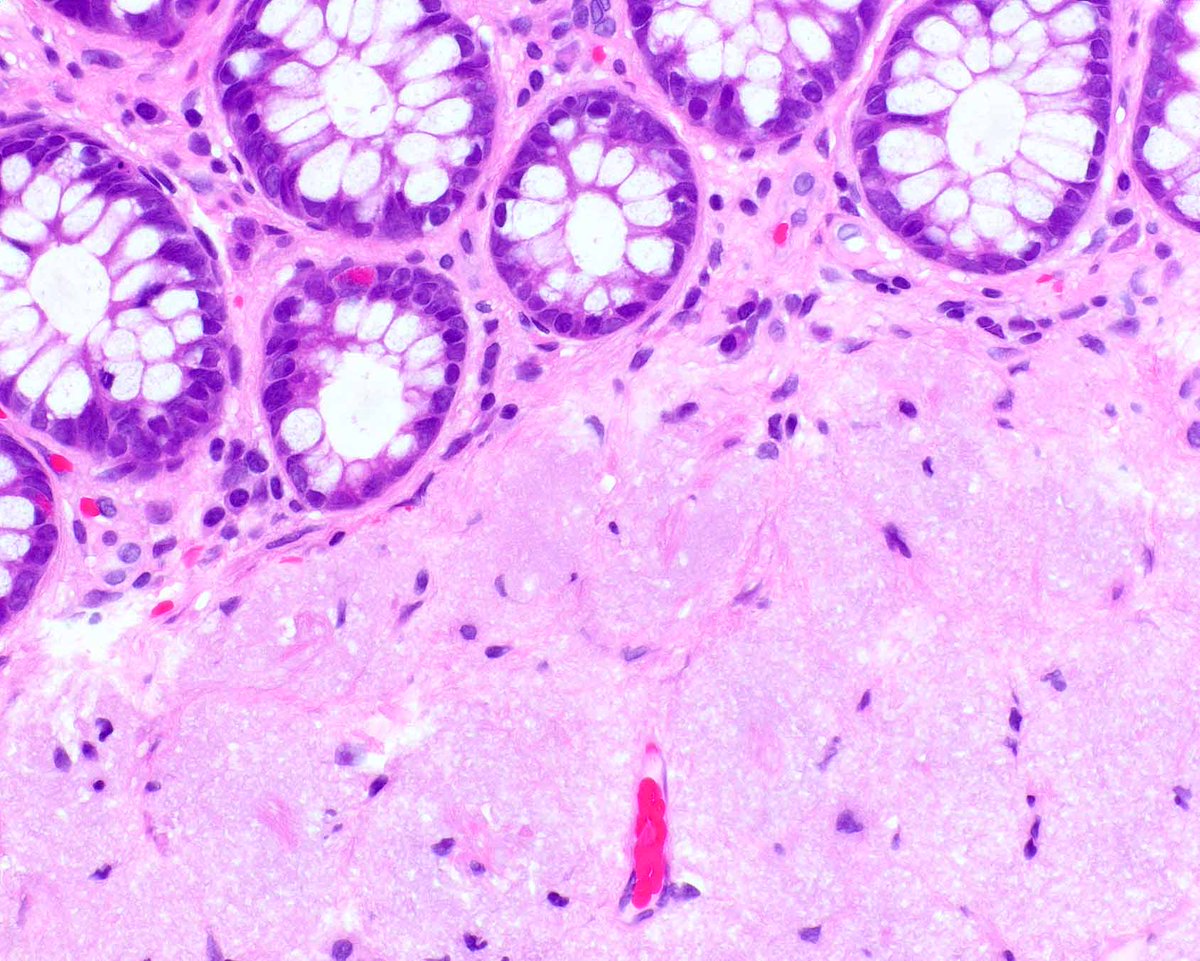
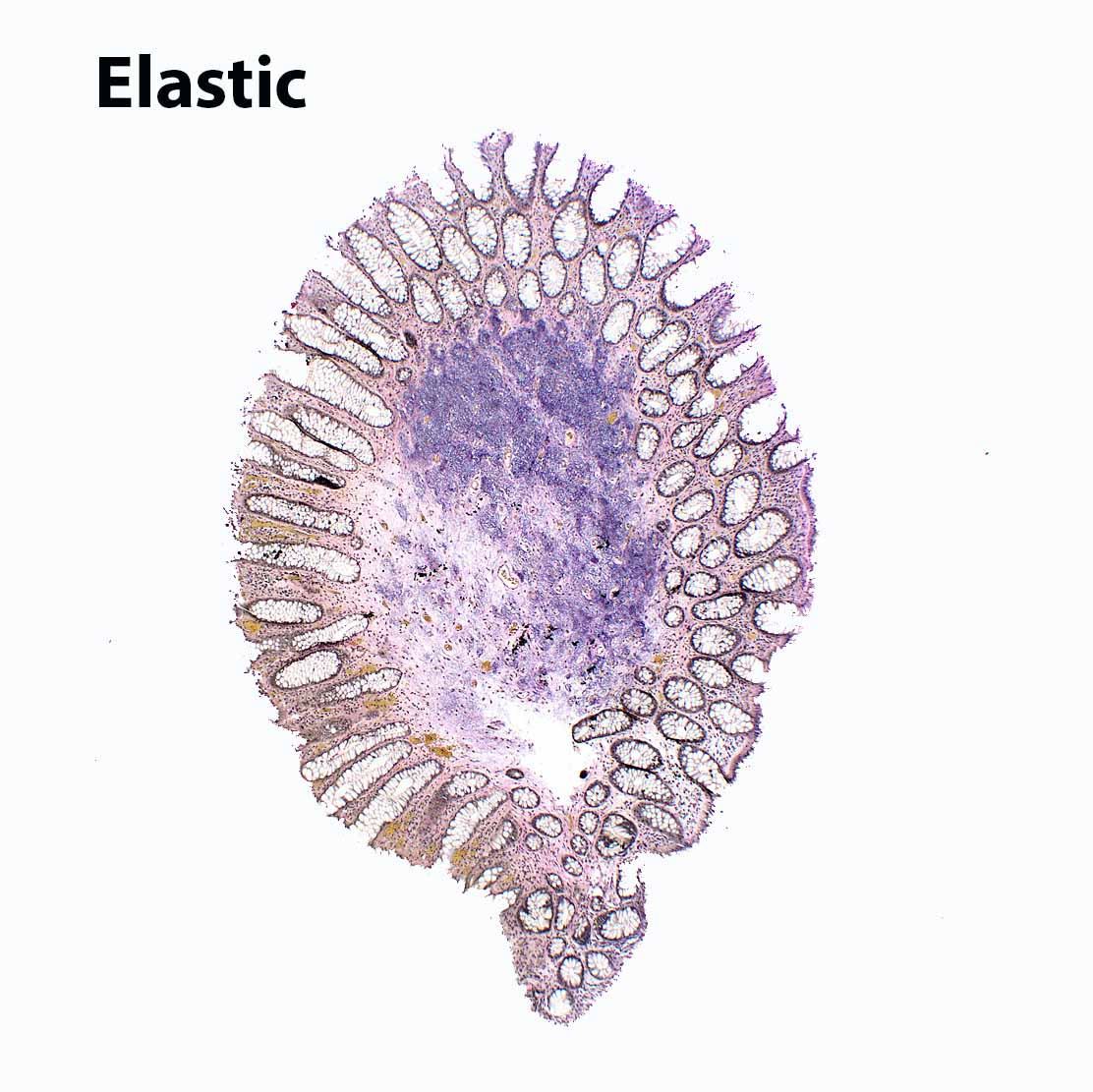
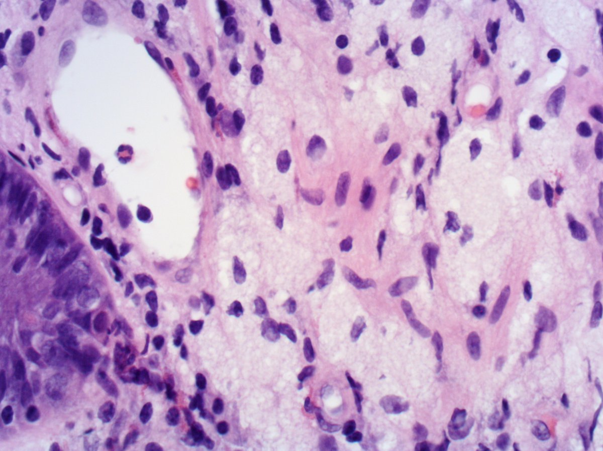
Intestinal elastosis/elastofibromatous change produces a polyp and probably reflects a damaged submucosal blood vessel.

5
1,217
23 Sep 2025
23 Sep 2025

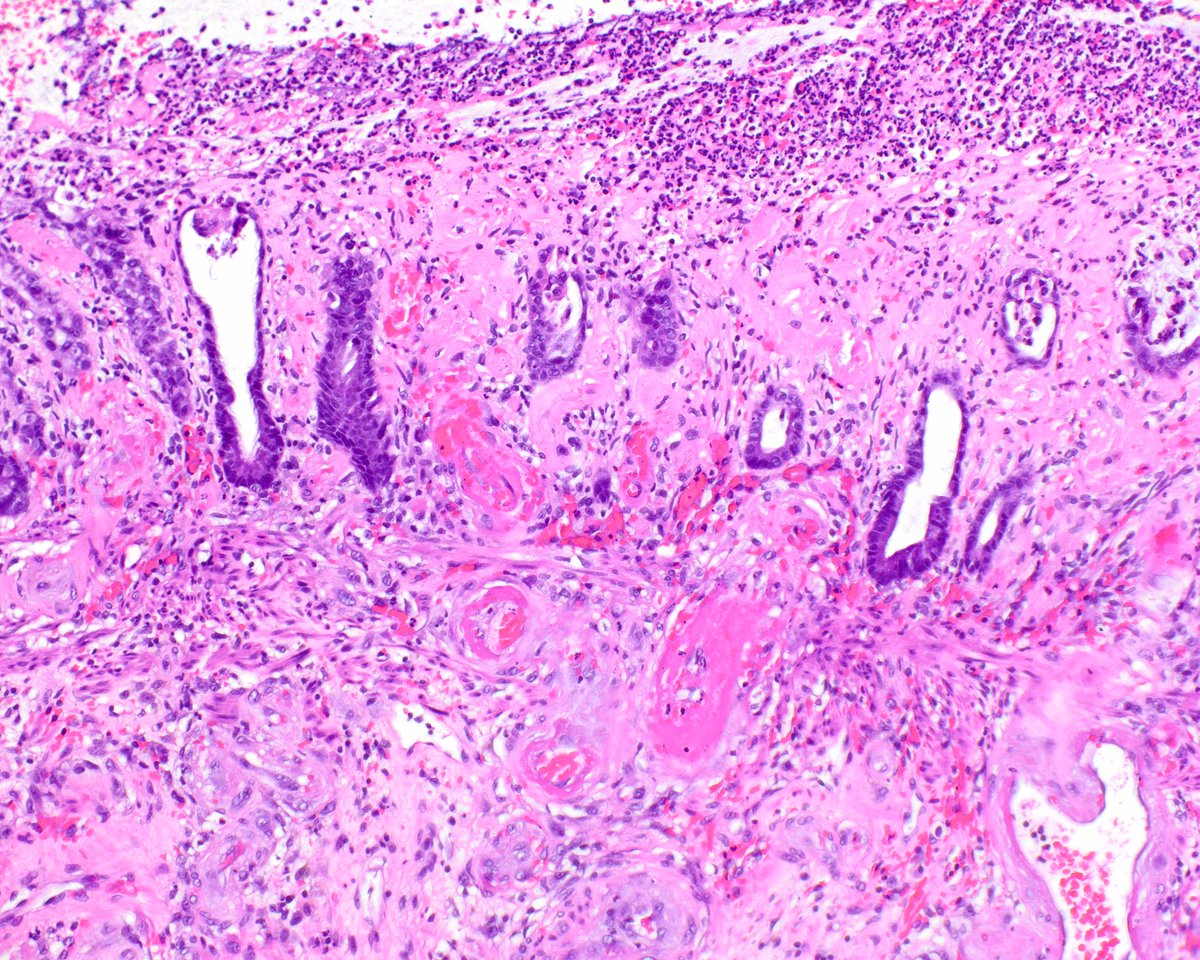
Appendicitis with an incidental serrated epithelial proliferation. The preferred terminology is "serrated polyp" rather than SSL as they have different genetics to their colonic counterparts. NB this is not LAMN as the muscularis mucosae is intact @Pathoutlines @RaulSGonzalezMD


1
3
25
2,381

29 Aug 2025
One of our talented PGY-2s, Mohamed Moustafa, presented a case report on a rare instance of GIST involving the ovary this past weekend at the Texas Society of Pathologists’ Summer Conference.
#PathX #PathTwitter #Pathology #GIPath #PathResidents #PathResEd

3
11
44
2,480
27 Aug 2025
Join us for our residency virtual open house on September 9th at 7 PM CST.
Zoom link for the Open House: utsouthwestern-edu.zoom.us/j…
#pathology #pathmatch2026 #pathresidents #PathResEd #pathtopath

1
8
16
6,912
31 Jul 2025
30 Jul 2025

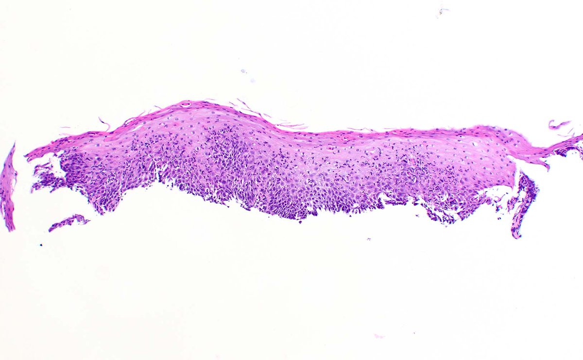
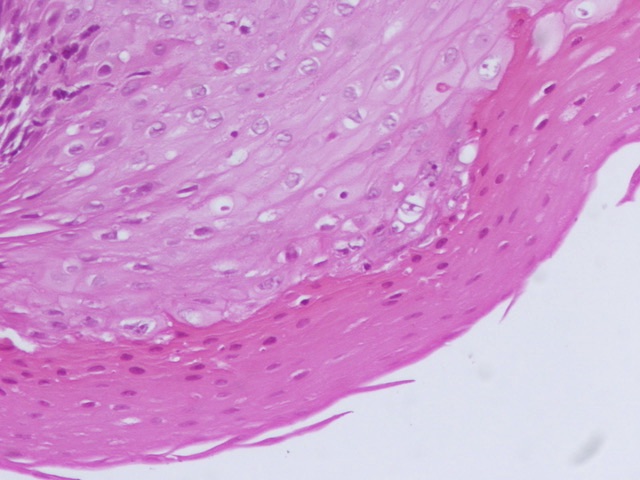
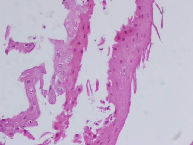
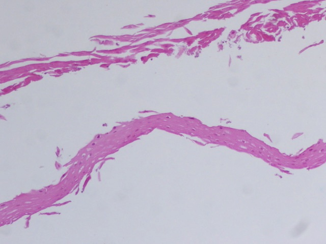
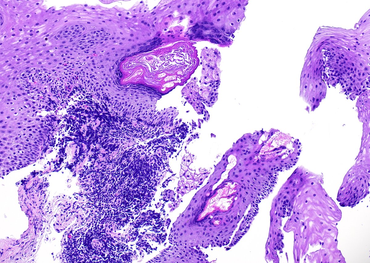
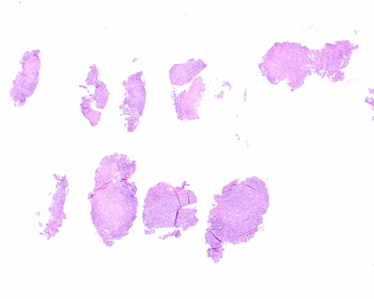
#GIPath #Diagnosethis #Pathresidents 1/2
Look what crossed my my microscope ?
The key is in the bitonal hue of this esophagus with sharply demarcated layers ,strong pink superficial ,intraepithelial cleft ,partially detached ,other fragments with just strips of superficial necrotic epithelium .
Make the Dx ,then go to the endoscopic appearance bonus .
#PathOutPic

1
4
650
30 Jul 2025
29 Jul 2025

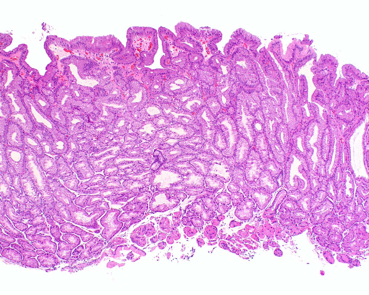
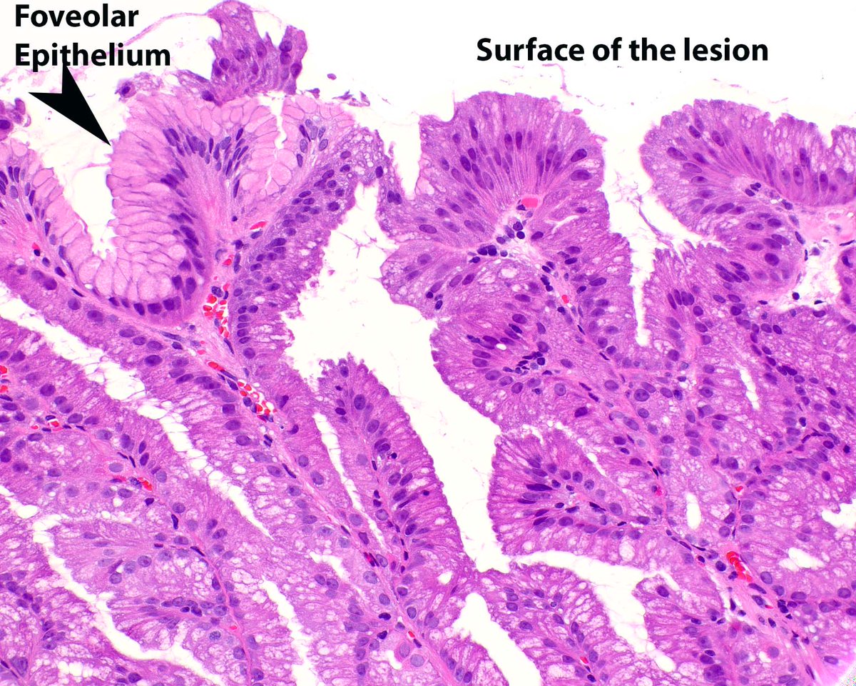
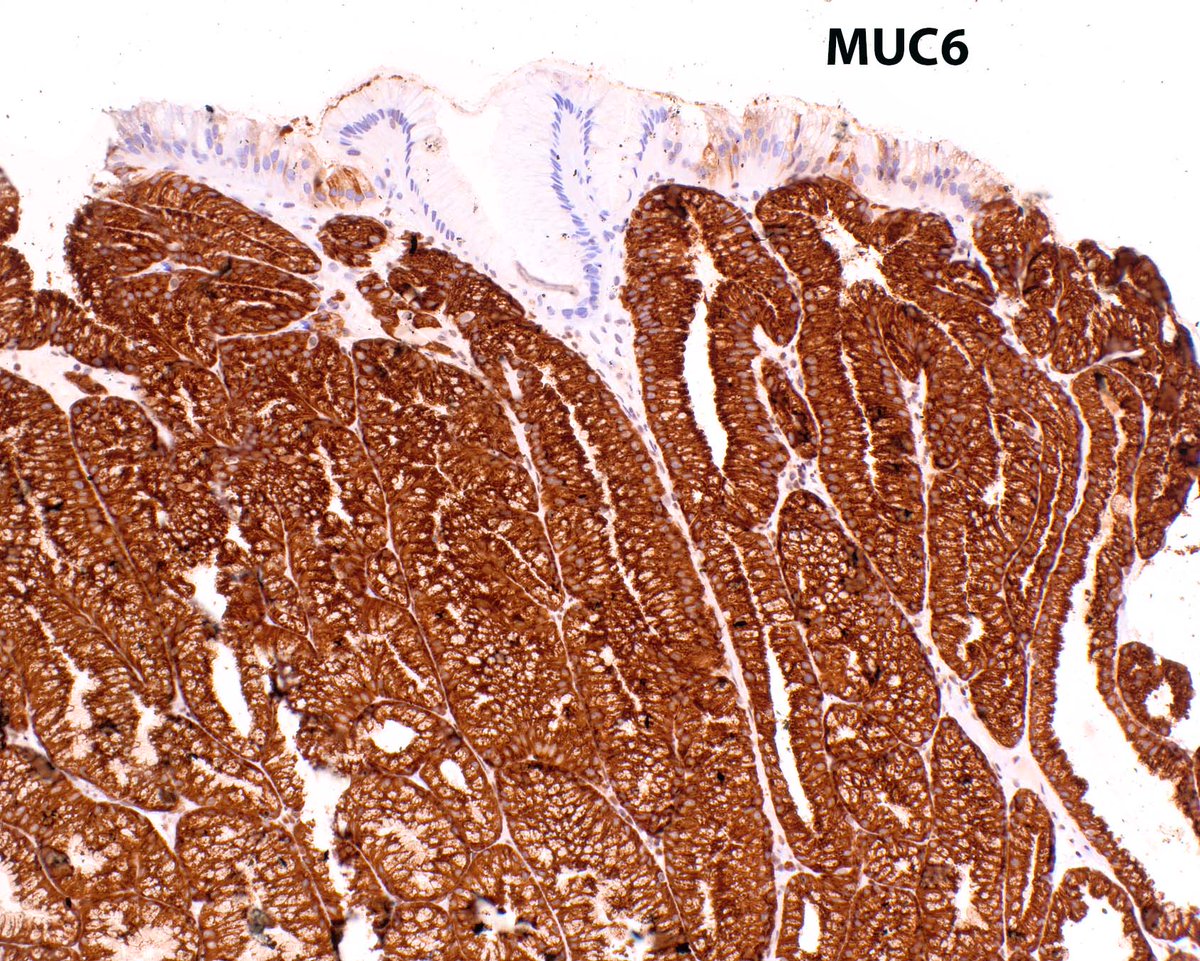
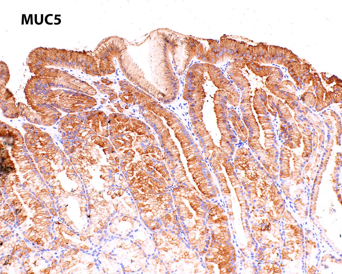
Pyloric gland adenoma in uninflamed gastric body. There is an association with autoimmune gastritis (pyloric metaplasia progressing to pyloric adenoma) but this patient does not have it. Note the appearance of the surface of the adenoma versus the adjoining foveolar epithelium.
4
424
4 Jul 2025
4 Jul 2025
Rectal muciphages; useful to identify the distal biopsy in an unlabelled colonic series. #Pathologists dont confuse them for sampling of a xanthoma! #GIPath


2
13
881
30 Jun 2025
🚨🚨🚨#GIpath pitfall. Beware 👀! 🚨🚨🚨
#pathology #pathX #PathTwitter #PathResEd #pathmatch2025 #pathmatch2026
4
16
64
4,336
12 Mar 2025
Excellent example of educational post
#PathTweetAward #PathResEd
@TheShorePath
@JuanPathMD
@LordoftheSlides
@Maicroanatomy
@ElaineHuMD
2
123
29 Jan 2025
Dr. Elisa Lin, PGY3, gave a talk to high school students about cancer and leukemia. You can now watch it here:
vimeo.com/1040162368/d28d699…
or youtu.be/nCv7tuNthzQ?si=Lqyq…
#POPSTARS #pathology #MedTwitter #PathX #PathTwitter #PathResEd
2
7
1,049
10 Jan 2025
8 Jan 2025

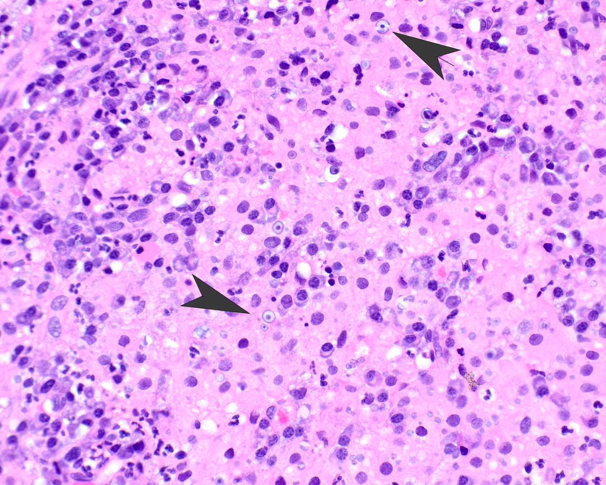
Help needed with interpretation of this mismatch repair pattern!
H&E of an upper GI adenocarcinoma 👇

1
3
498
9 Jan 2025
9 Jan 2025


1
1
10
673
16 Aug 2024
15 Aug 2024
This example of colorectal malakoplakia presented as a mass lesion. The pale pink appearance of the histiocytes is a clue to search for Michaelis-Gutmann bodies. #umiamipath
Reference: Am J Surg Pathol. 2020 Sep;44(9):1251-1258. PMID: 32301754.
3
278
13 Aug 2024
Join us for our virtual open house on Wednesday (8/28) at 7 PM.
Zoom link for the Open House:
utsouthwestern-edu.zoom.us/j…
#pathology #pathmatch2025 #PathResEd

1
15
33
5,625
30 Jul 2024
30 Jul 2024
Myointimal hyperplasia of mesenteric veins has been known for decades to cause segmental ischemic disease-cured by removing the affected segment. More recently, @RhondaYantiss did a wonderful job describing the findings in mucosal biopsies. #UMiamiPath PMID: 28817406


1
4
401