The #SNMMI 2023 Mid-Winter & ACNM Annual Meeting kicked off this morning with a "New Image Interpretation Training" session, where participants learned how to accurately interpret new diagnostic agents and identify quality improvement opportunities when delivering patient care.
Interested in getting involved with @ACRRFS? Apply for the Executive Committee and Nominating Committee! It’s an incredible opportunity to influence policy and develop resources for #RadRes and #RadFellows. Cultivate leadership skills and gain lifelong friends along the way!
What a great start to the ACNM/@SNM_MI Hot Topics Webinar Series. Nothing gets hotter than #PSMA right now. It was a privilege to have Dr. Steven Rowe @HopkinsNucs speak on PSMA PET. Excited for the amazing line-up of talks ahead.
#nucmed#radtwitter#FOAMed
A novel nuclear imaging probe can measure gastric cancer patients' response to treatment within days, a potential breakthrough for treatment-resistant cases. Read about research by @ShadiEsfahani using a 2018 RSNA Research Resident Grant. bit.ly/3r59LzB#RandEFoundation
**CALL TO ACTION** CMS is proceeding with changes to the MPFS that mean SIGNIFICANT CUTS for #NuclearMedicine (9% or more). SNMMI and 200 other societies are urging Congress to help. Please SUPPORT that message--write your members of Congress TODAY at ow.ly/M82M30rXrTO.
What's the buzz with Fibroblast activation protein? 68Ga-FAPI is the new radiotracer on the block that's challenging the likes of 18F-FDG. How does FAPI PET perform in breast cancer? - see for urself in this #RadInTraining#InAMinute
@RadITrainEditor @RSNA@radiology_rsna
A 2-year life-changing postdoctoral training experience with the incredible Dr. Julie Price (T32 Postdoc)! Training in multimodal neuroimaging (PET/MR) focusing on key research areas in Alzheimer’s disease and related dementias
Please share widely! #ENDALZpricelab.martinos.org/t32-ad…
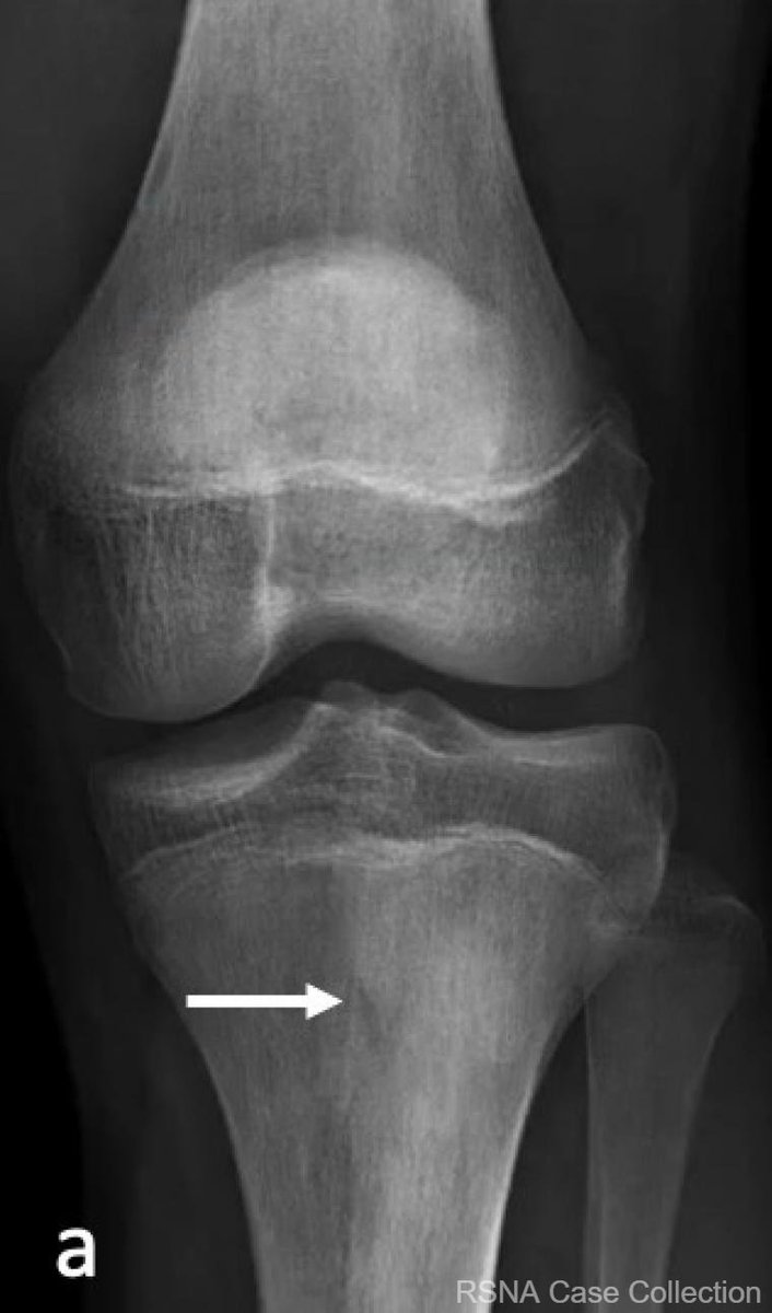
ALT Anteroposterior radiograph of the left knee demonstrates subtle, ill-defined sclerosis in the left proximal tibial metadiaphysis (arrow).
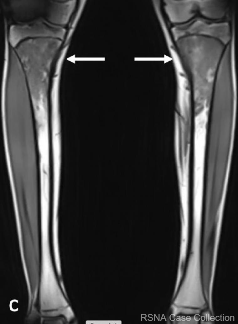
ALT Coronal T1-weighted image of the bilateral tibias demonstrate symmetrical T1 hypointensity involving the bilateral, proximal tibial metadiaphyses.
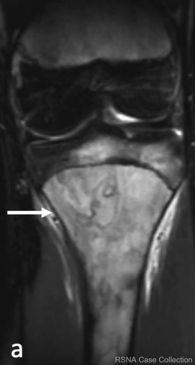
ALT Coronal STIR image of the left knee demonstrates confluent hyperintense intramedullary signal involving the left proximal tibial metadiaphysis and tibial epiphysis. There is a serpiginous area in the medullary bone of the left medial tibial metaphysis (arrow) which may represent a bone infarct.
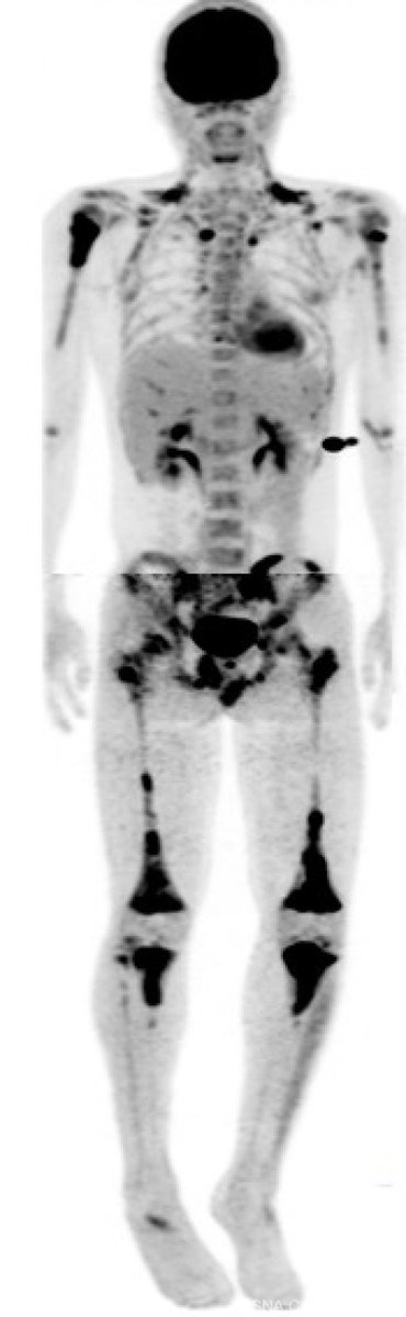
ALT Maximum intensity projection (MIP) image from a whole-body PET/CT demonstrates extensive multifocal hypermetabolic intramedullary lesions in the appendicular greater than axial skeleton.
#SNMMI21 Late-Breaking Session Panelists Added! “PSMA Radioligand Therapy: Updates from the VISION Trial.” The Phase 3 #VISIONTrial is evaluating the efficacy of 177Lu-PSMA-617 (LuPSMA) in the treatment of patients with PSMA-positive metastatic castrate-resistant prostate cancer.
Thomas J. Ruth, PhD, known for his contributions to nuclear medicine and nuclear chemistry, has been named as this year’s recipient of the SNMMI Georg Charles de Hevesy Nuclear Pioneer Award. Congratulations Dr. Ruth! #SNMMI21#NuclearMedicine
I'd like to congratulate @ShadiEsfahani, whose Research Seed Grant application was among @RSNA's top-ranked and approved for funding by the R&E Board of Trustees. Well done! #MGH#Radiology