
Part of the University of @Penn : cellular & molecular aspects of the brain; systems & computational neuroscience; behavior & cognition; & pathology
- Tweets 942
- Following 323
- Followers 685
- Likes 428


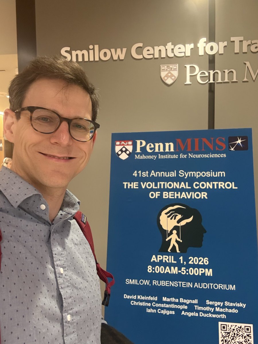
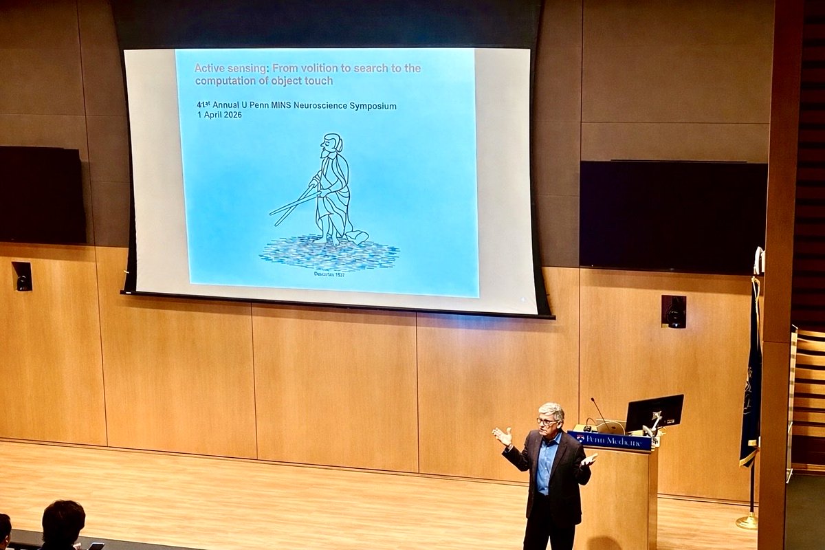
ALT Sergey in front of a sign advertising the event
ALT Formally set dining table with a city view of Philadelphia


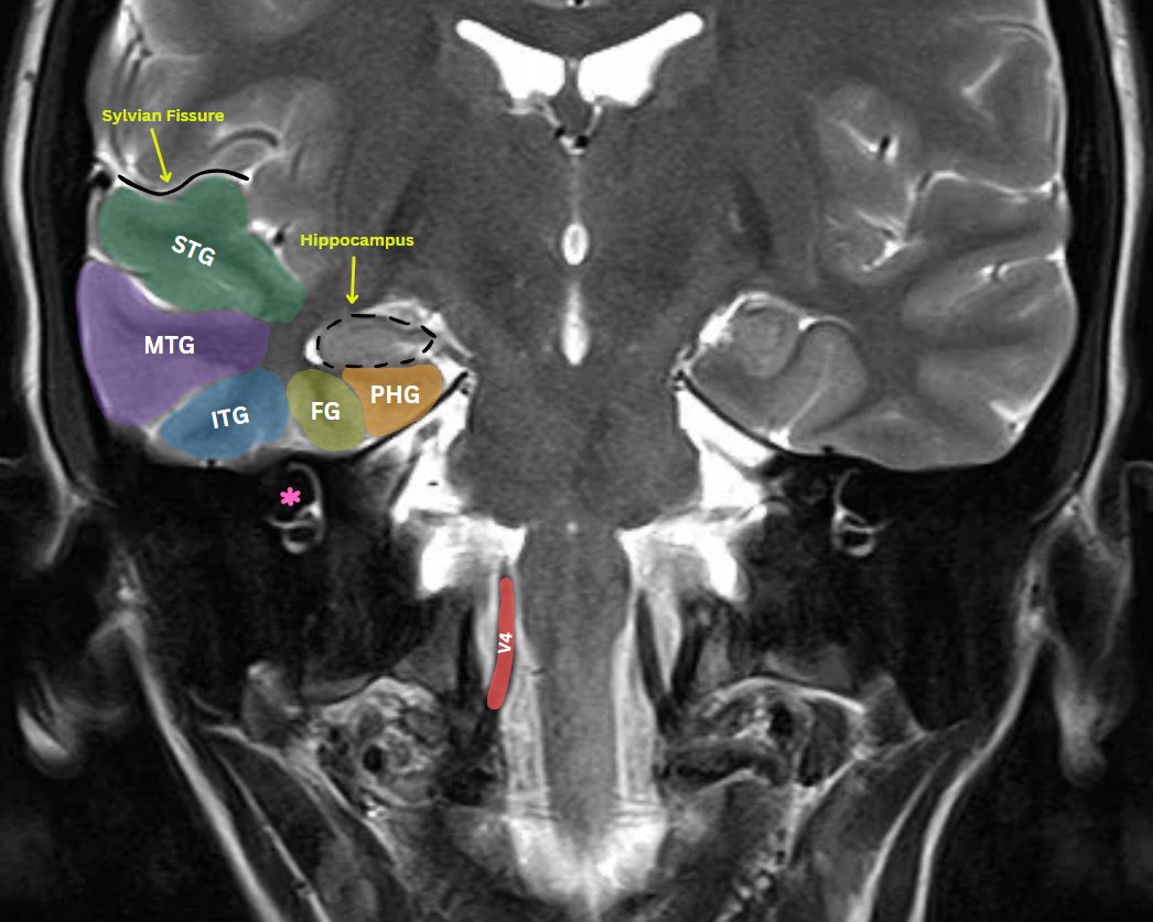


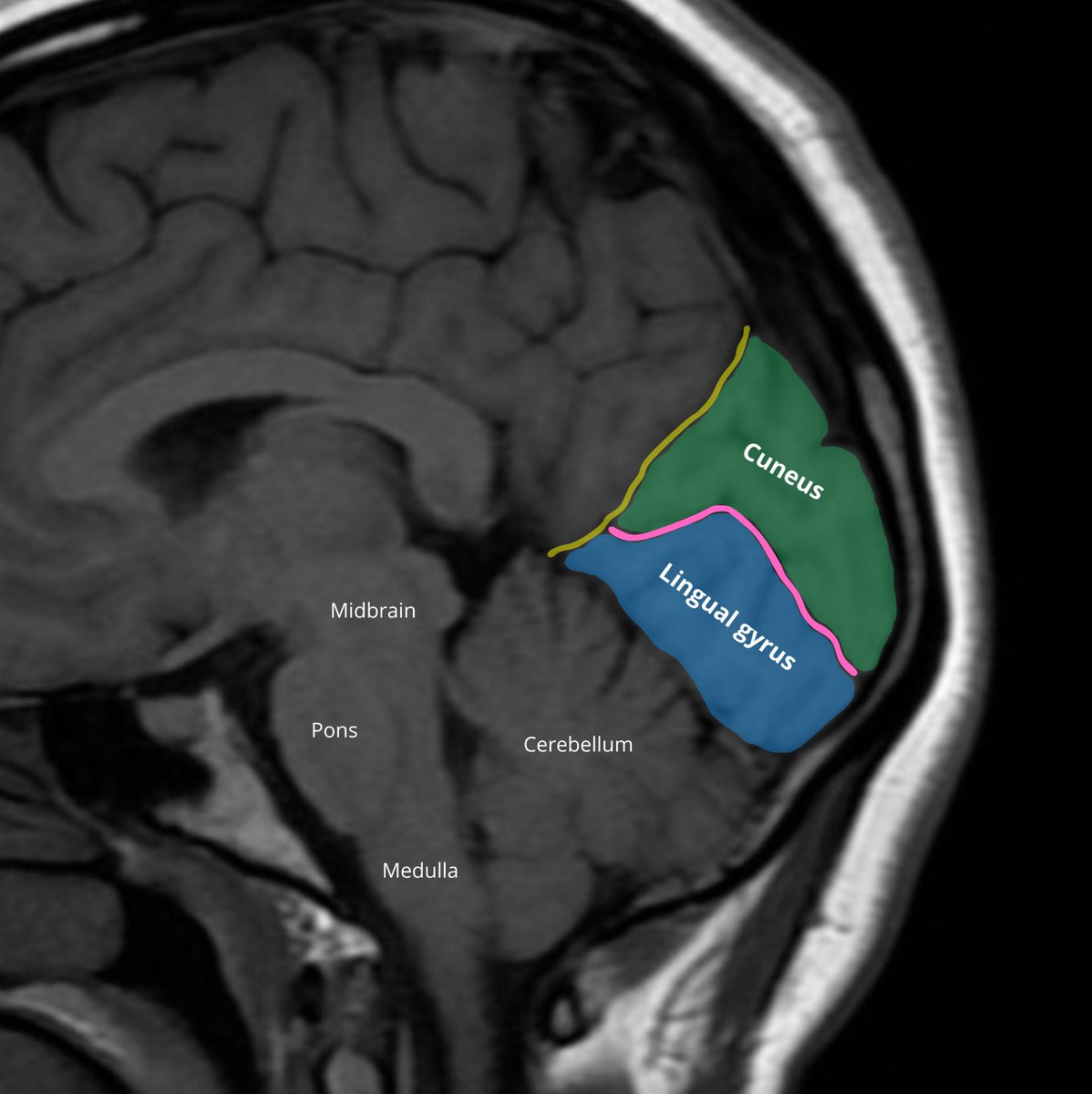
ALT Image annotation: Raisa Amiruddin and Nikolay Yordanov Source of the native MRI image: Gaillard F Normal brain (MRI). Case study, Radiopaedia.org (Accessed on 26 Jan 2025) https://doi.org/10.53347/rID-37605

ALT Image annotation: Raisa Amiruddin and Nikolay Yordanov Source of the native MRI image: Abidin M, Normal MRI Brain. Case study, Radiopaedia.org (Accessed on 18 Nov 2024) https://doi.org/10.53347/rID-153576


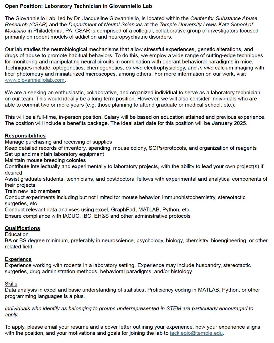
ALT Job posting for a laboratory technician in the Giovanniello Lab at Temple University.