
We develop microscopy methods to observe life on the nanoscale
Joined December 2015
- Tweets 249
- Following 292
- Followers 1,035
- Likes 1,184
36 Photos and videos


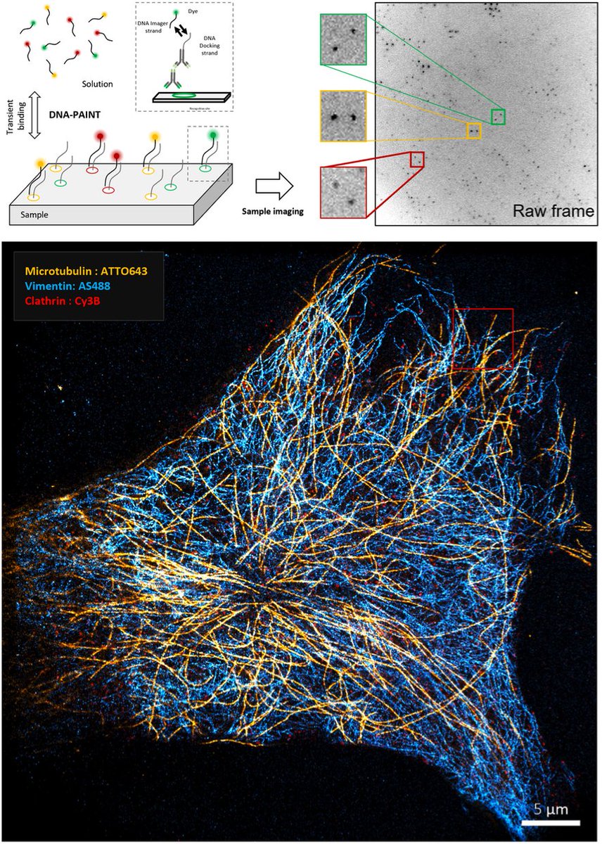
Gen-AI producing fancy data augmentation for Super-resolution microscopy! Great work @AlonSaguy and team! @HenriquesLab @HeilemannLab
18 Oct 2024

If robots could dream of microtubules, how would they look like? An amazing story by Alon Saguy, @ShechtmanLab and colleagues. Proud we could contribute to it
onlinelibrary.wiley.com/doi/…

1
3
9
1,777
Great work @yevgenin @DanielleSapir et al.!
21 Jun 2024

Thrilled to share OM2Seq! We applied #AI methods to #Genomics, improving the speed and accuracy of optical genome mapping for fast diagnostics of diseases. @DanielleSapir, Tahir Detinis Zur, Nir Weinberger, @boknilev, @hagenom, @ShechtmanLab
🔬🚀
1
311
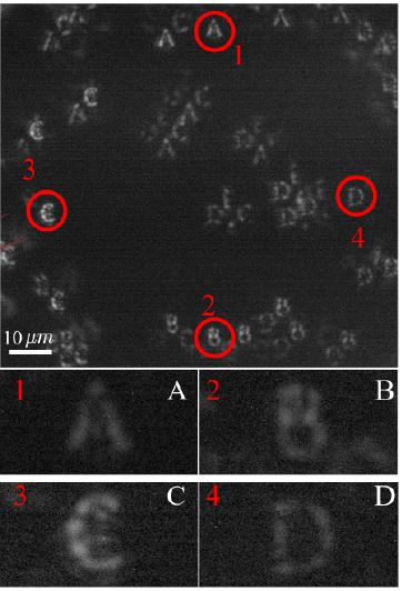
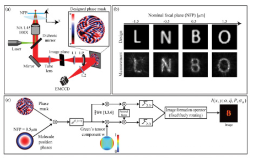
PSF engineering typically requires a 4-f system, but this is not always feasible, e.g. due to size constraints. Nadav Opatovski attached a phase mask directly to the objective lens, see beads defocusing here. Tetrapod mask shown here (wait for the end):
1
3
15
2,018
This enables PSF engineering in space-limited microscopy systems, e.g. that fit into incubators. Uses include extended depth-of-field and snapshot 3D spheroid from 2D, using algorithms by @EliasNehme9. Bonus: Obtaining training images is easy with a high-throughput microscope.
1
2
735
We used the Incucyte system by @SartoriusGlobal. All this and more in our latest paper: link.springer.com/article/10…. Big thanks and congrats to all co-authors and collaborators: Nadav, @EliasNehme9 Noam, Ilana, Reut, Boris, Paul, @AlaloufOnit , @SartoriusGlobal !
4
291
🥳 DBlink is now peer reviewed and improved! Online in @naturemethods: rdcu.be/dhUmZ
Congrats @AlonSaguy, @AlaloufOnit, @HeilemannLab et al.!

New prepint just out! Live cell single molecule localization microscopy at >3 fps. The information is mostly there - just spread out in space and time :)
We use a recurrent neural net (LSTM) some assumptions to extract it. All details here:
biorxiv.org/content/10.1101/…
5
12
58
8,683
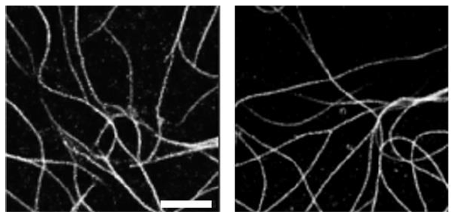
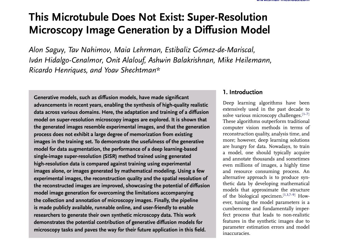
One of these super-resolution microtubule images was captured by a microscope and the other one was generated by a diffusion model. Can you tell which is which? Try more here: clever-microscopy.com/this-m…
1/4
3
3
23
6,428
Alon Saguy et al. trained a diffusion model to generate synthetic images of super-resolution microscopy images using data of only a few images from ShareLoc (shareloc.xyz).
2/4
2
2
625
More info in the preprint: biorxiv.org/content/10.1101/………, where we demonstrate the use-case of generating training data for CARE (by
@martweig et al.)
3/4
315
This is a very ongoing project, with many open questions: what are the performance limits? How much can we really gain beyond the information in the training data? and of course: what would be the best uses of something like this in super-resolution microscopy?
4/4
216
Great work by @yevgenin applying information theory to optical genome mapping, with implications to optimal label design
29 May 2023
New #preprint!
TLDR: Applying #informationtheory on #OpticalGenomeMapping🧬, one can choose pattern-labelling #enzymes giving #10x accuracy! 🤯
biorxiv.org/content/10.1101/…
To illustrate, let’s play a game! ⬇️ (1/4)
1
5
642
Nano Bio Optics Lab (Yoav Shechtman) retweeted

5 Apr 2023
Major paper announcement:
Review “Fluorescence Microscopy: a statistics-optics perspective” is now on arxiv.
with Fazel, Grussmayer, Feldman, @LabRadenovic, @ShechtmanLab, @JoergEnderlein
Add exercises and turn into textbook? 😜
arxiv.org/abs/2304.01456
#FOM2023 #microscopy
1
15
45
9,629
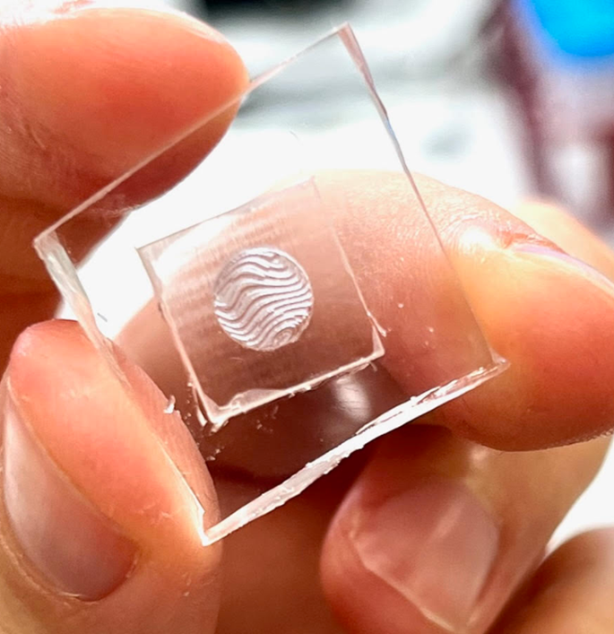
Computational microscopy often relies on some kind of wavefront shaping by diffractive optical components, which can be very difficult to fabricate.
This motivated us to utilize 3D printing near index matching for simple & cheap DOE fabrication.
arxiv.org/abs/2303.15197
1
7
25
5,726
We generate a variety of solid elements, including microlenses, vortex plates for MINSTED and more, and pretty challenging PSF engineering masks! Thanks to our collaborating groups, Ady Arie, @Stefan_W_Hell @AleksPonjavic, in this multi-national effort led by Reut Kedem Orange!
1
6
453
2 bonus points if you can guess what this DOE does (clue in the preprint 🙂)
1
1
6
562
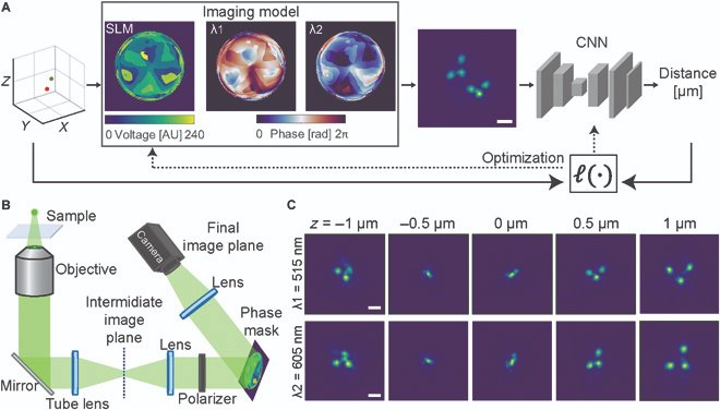
In many (most?) cases in fluorescence microscopy, in which the positions of spots are determined, it is not really their global positions that are of interest, but rather their *relative* positions, i.e. their distances from one another.
23 Dec 2022

#IntelligentComputing New research article
Learning Optimal Multicolor PSF Design for 3D Pairwise Distance Estimation
Ofri Goldenberg, Boris Ferdman, Elias Nehme, Yael Shalev Ezra, and Yoav Shechtman @ShechtmanLab
Special Issue "Computational Imaging"
spj.science.org/doi/10.34133…
1
2
13
2,061
Consider the problem of determining the 3D distance between a pair of 2-colored spots. One option is to find x1,y1,z1 and x2,y2,z2, e.g. using a 3D PSF, and calculate the distance. But why not optically encode the *distance*, rather than individual positions?
2
4
449
Just out: Ofri Goldenberg learned and implemented a multicolor phase mask decoding net for determining the distance between a pair of 2-colored spots in 3D. The net outputs the scalar distance of interest. spj.science.org/doi/10.34133…
253
cool idea implemented using simple elements!

Doing multicolor (single-molecule) imaging? Tired of waiting for each color?
Want simultaneous imaging with full field of view but without the multicamera hassle?
Take a look at our 'Circulator'! 🔥💪
t.ly/lHZq. Very excited so please RT! (1 of 5)
#microscopy
1
1
3
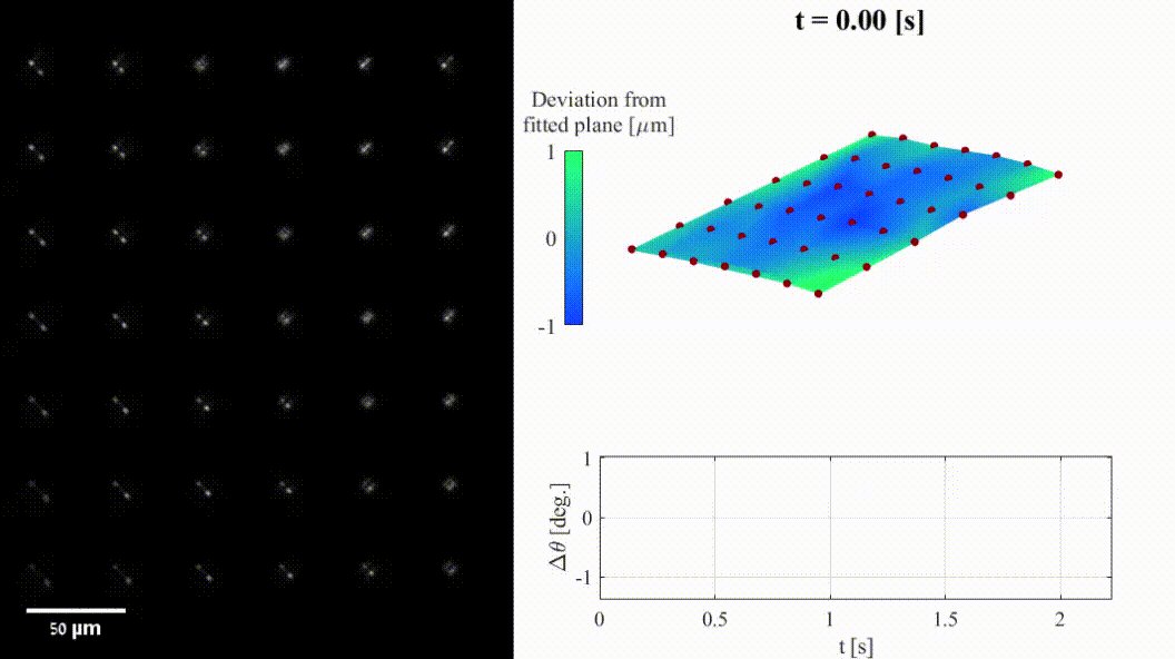
Can you determine your distance from a truck the same way you determine the z position of a fluorescent molecule? Of course you can, all you need is a PSF-engineered telescope! Adventurous Nadav Opatovski built one: doi.org/10.1364/OE.472150
2
9
46
As @HenriquesLab noted in the last SMLMS regarding the blinking Eiffel tower - Maxwell's equations are the same regardless of scale...
BTW the failure of the active rangefinder in the gif is due to scatter from dust from the truck; PSF engineering is much more robust to it.
1
1
4
Phase mask is a double-helix, and for image analysis we used @SjoerdStallinga's excellent cepstrum method or a neural net by Dafei Xiao. More info in the paper realization how fortunate we are in microscopy to neglect atmospheric turbulence an excuse for us to buy a drone.
1
2