
Co-founder and CEO of Viventis Microscopy
Joined September 2012
- Tweets 57
- Following 35
- Followers 101
- Likes 17
Photos and videos
Petr Strnad retweeted

13 Jun 2024
Interested in extracting information from movies like this? Have some programming background? We have an open JRF position in my lab symmetry-lab.com
Check out the ad
main.tifr.res.in/maincampus/…
Pass this around if you know of someone who fits the requirement
Deadline June 26
1
36
62
14,636
Petr Strnad retweeted

11 Jun 2024
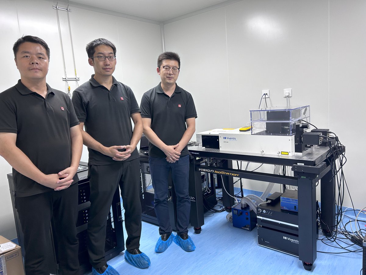
🎥 Researchers at the FMI and @ViventisMicro teamed up to develop a cutting-edge light-sheet microscope that has the potential to transform imaging studies and enable scientific breakthroughs.
@priscaliberali @FranziskaMoos @SimonSuppinger @TsiairisLab
8
20
2,555
Petr Strnad retweeted

📣 Big News! Viventis Microscopy is now part of @LeicaMicro
The Viventis LS2 Live microscope is available globally from today. Discover detailed volumetric imaging to explore life in its entirety.
Press release 👉 fcld.ly/w9s7z3d
#LightSheetMicroscopy #liveimaging
4
16
71
8,134
Petr Strnad retweeted

7 May 2024
📣 Exciting news! We have integrated cutting-edge light sheet technology from Viventis Microscopy @ViventisMicro into our portfolio. The Viventis LS2 Live microscope enables volumetric imaging to explore life in its entirety.
#LightSheetMicroscopy #liveimaging
1
7
21
2,849
Petr Strnad retweeted
We are happy to see the concept behind our LS2 Live #lightsheet microscope published in @naturemethods
nature.com/articles/s41592-0…
#organoids #liveimaging #microscopy
1
6
33
2,413
Petr Strnad retweeted
We would like to close this year by sharing two freshly released studies that use our #lighsheet system:
1) The @KenzoIvanovitch lab @ucl uses live cell imaging during mammalian gastrulation to reveal the origin of cardiac progenitors.
biorxiv.org/content/10.1101/…
1
8
40
2,536
Petr Strnad retweeted
The latest work from @OatesLab @EPFL_en shows how cells are instructed from the segmentation clock to form somite boundaries in #zebrafish embryo.
biorxiv.org/content/10.1101/…
Check out this wonderful movie acquired on our #lightsheet by lead author @Olivier_Venzin.
6
59
5,566
Petr Strnad retweeted
We are happy to announce that our LS2 Live #lightsheet is now available in China 🇨🇳 thanks to our partner Quantum Design China.
We also would like to celebrate the first successful installation of our system at the National Institute of Biological Sciences (NIBS), Beijing.
1
2
6
939
Petr Strnad retweeted

2 Oct 2023
I am excited to show you these beautiful movies! A great effort together with @TweetPetrStrnad at @ViventisMicro to design a new #lightsheet for long term high throughput live imaging of large specimens #organoids, #embryoids and entire animals #hydra. dlvr.it/Swnqmw
6
66
328
46,871
Petr Strnad retweeted
Impressive work on live #imaging of brain #organoids using our #lightsheet system from @GrayCampLab and @TreutleinLab.
This is a huge effort from @Jain_Akanksha_ and @GillesGut both in microscopy and image analysis. Many Congratulation !!
28 Aug 2023

I am very excited to share my postdoc work “Morphodynamics of human early brain organoid development”, where we take a multiscale morphodynamic view of human brain organoid development. In collaboration with @GillesGut @GrayCampLab @TreutleinLab biorxiv.org/content/10.1101/…🧵(1/9)
4
19
2,640
Petr Strnad retweeted
The beautiful work from @diane_pelzer in @maitrejl Maître's lab in now published in @emboJournal.
Our LS1 Live #lightsheet was instrumental to investigate the mechanism behind cell fragmentation in pre-implantation #embryos.
embopress.org/doi/full/10.15…
1
5
16
1,714
Petr Strnad retweeted
Check out the last preprint from @Tanaxolotl Tanaka’s group. @teresakatharin1 and colleagues used our LS1 microscope to show how neural tube organoids form their ventral floorplate!
Read the thread below for more information about the mechanism
#microscopy #organoids #lightsheet
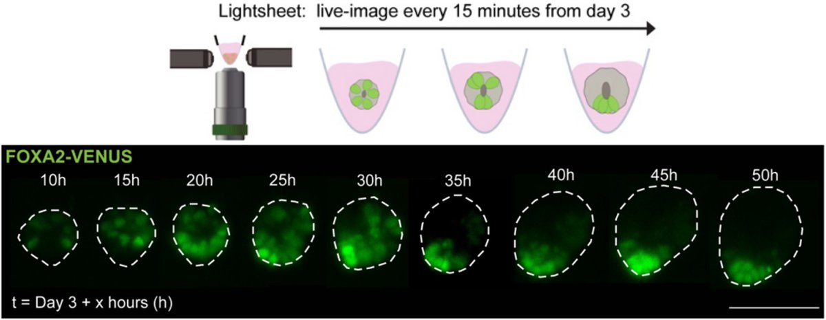
ALT Top: schematic (created with BioRender) of timelapse imaging of double reporter on the Viventis lightsheet microscope. Full image stacks with 3 µm z step size were taken every 15 mins starting 6 hours post RA removal on Day 3 for 48-75 hours. Bottom: images from timelapse series of a NTO (maximum intensity z-projections) following the spatial organisation of FOXA2 (green) over 50 hours showing self-organisation from a dispersed (“salt-and-pepper”) into a clustered state. Dashed lines mark the outlines of the NTO. Scale bar, 100 µm.
28 Jun 2023

Have you ever wondered who organizes the organizer? Excited to share our latest discoveries about how a global application of retinoic acid (RA) can induce polarized neural tube organoids (NTOs).
For more details, check out our pre-print below:
biorxiv.org/content/10.1101/…
1/6
5
13
2,029
Petr Strnad retweeted
We are very happy to have Davide Gambarotto (@DavideGambarot2) joining the team as application specialist.
Davide has worked with expansion-super resolution techniques and has extensive experience in imaging and cell biology applications.
#microscopy #organoids #lightsheet
1
1
17
1,516
Petr Strnad retweeted
Next week we will be attending the @BaCell3D on May 8-9 in #Basel.
Come and talk to us to know more about our latest LS2 Live system and how we image organoids for extended period of time using #lightsheet.
Video credit @FranziskaMoos, @priscaliberali lab.
6
29
2,087
Petr Strnad retweeted
Our CEO Petr Strnad (@TweetPetrStrnad) is presenting the technology behind our new LS2 Live at #FOM2023.
You can also discuss the several imaging applications with our collaborators Gustavo @gusqgmed and @FranziskaMoos from @FMIscience.
#Organoids #Imaging #LightSheet
5
29
2,459

31 Jan 2023
Our company released a new microscope system! A milestone for @ViventisMicro and an amazing journey for our team. We've put years of work, energy and enthusiasm into creating an instrument that we hope will help scientists make discoveries. Thank you to everyone who contributed!

🔬 New product launch 🔬
Today, we are proud to launch our new #LS2Live!
With our new product, you can image, larger & more sensitive samples like #organoids and #embryos in a multiplex and parallelized fashion.
1/2
9
27
3,881
Petr Strnad retweeted
🔬 New product launch 🔬
Today, we are proud to launch our new #LS2Live!
With our new product, you can image, larger & more sensitive samples like #organoids and #embryos in a multiplex and parallelized fashion.
1/2
1
15
35
16,626
Petr Strnad retweeted
We were excited to see this timelapse from Sera Weevers at @TsiairisLab, @FMIscience.
In collaboration with @priscaliberali's lab, they used one of our #lightsheet #microscopes to show development of 3 Hydras.
Stay tuned to see how this large & light sensitive sample was imaged!
1
12
36
Petr Strnad retweeted
@IshiharaKsk, Arghyadip Mukherjee & collaborators using brain tissue #organoids and our #LS1 Live instrument have measured, described & manipulated the physical mechanisms that lead to the emergence of shape & architecture in developing tissues!
go.nature.com/3tXDe0a @mpicbg
1
1
9
Petr Strnad retweeted
We are excited to extend our collaboration with @priscaliberali's lab and our fellow collaborators at @FMIScience to develop #lightsheet #imaging solutions for #organoids and #3DCultures - @gusqgmed and Franziska Moss.
We will update you with developments from the collaboration!
8
39