

🔬 April 20, 1940: The Day We Saw the Invisible 🔬
On this day 86 years ago, the first U.S. electron microscope was publicly demonstrated in Philadelphia, PA—and science would never be the same. 🧵⚡
The mastermind? Vladimir Zworykin—a Russian-American inventor working for RCA (Radio Corporation of America). 🇷🇺➡️🇺🇸
While traditional microscopes maxed out at ~1,500x magnification (limited by light's wavelength), Zworykin's electron microscope shattered that barrier—using electrons instead of light beams. 💡➡️🔬
The results were staggering: ✨
• Magnification up to 100,000x
• Resolution down to 2.5 nanometers
• Viruses—never before seen—suddenly became VISIBLE 🦠
This wasn't just an upgrade. It was a quantum leap into the nanoworld. 🌌
From that moment, scientists could:
• Study virus structures directly 🦠
• Analyze crystalline materials at atomic scale 💎
• Pioneer modern materials science & semiconductor research 🖥️
• Launch the field of nanotechnology 🧪
Zworykin (1888–1982) was no one-hit wonder. He also invented:
📺 The iconoscope—an early television camera tube
📡 Key components of modern TV broadcasting
But the electron microscope? That was his gift to every future biologist, chemist, and physicist. 🎁
The Philadelphia demo in 1940 marked America's arrival as a leader in high-resolution imaging—and opened the door to understanding life at its most fundamental scale. 🧬
Today, cryo-electron microscopy (cryo-EM)—a direct descendant of Zworykin's machine—wins Nobel Prizes 🏆 and maps proteins atom by atom.
All because on one April day, in one city, one inventor showed us how to see the invisible. 👁️✨
#HistoryOfScience #ElectronMicroscope #VladimirZworykin #RCA #Philadelphia #Microscopy #Nanotechnology #STEMhistory #Physics #Biology #Virology #MaterialsScience #ThisDayInHistory #April20 #ScienceFacts #Innovation #InvisibleWorld @RCA @NobelPrize @ScienceMag @Nature @PhysicsToday @ASM_Microbe @MicroscopyToday @PhillyHistory @AmerChemSociety @IEEEorg @BiophysicalSoc @Nanowerk @VirologyBlog

25
31
191


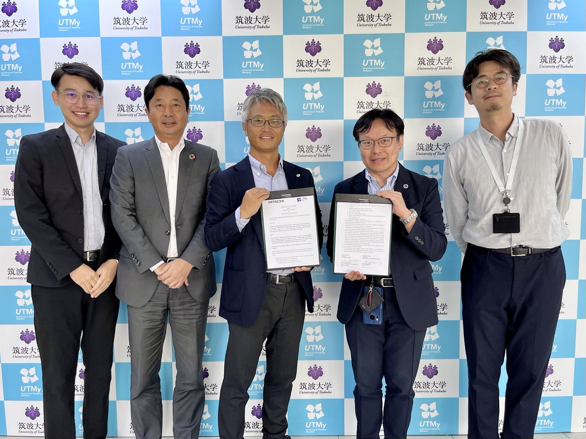
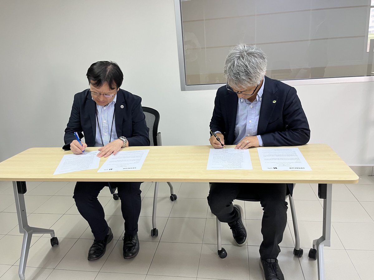
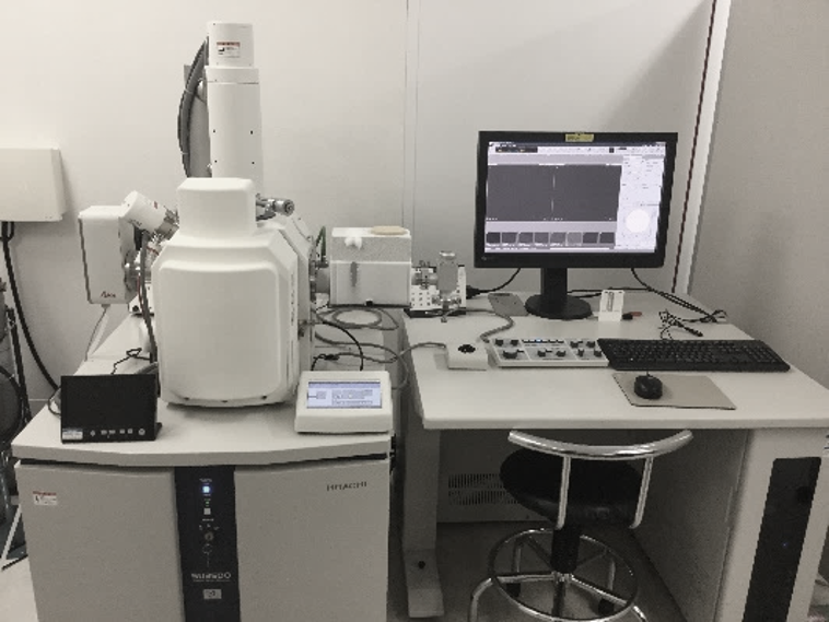
Exploring the Future of Electron Microscopy for Research
Hitachi High-Tech IPC (Malaysia) Sdn. Bhd. and University of Tsukuba, Malaysia have signed a Memorandum of Understanding (MOU) to explore and develop research needs in the field of Electron Microscopy. This collaboration marks the beginning of a long-term joint research initiative aimed at understanding how electron microscopy is currently used across the region and where the next frontier of research may lie.
Electron microscopy is an enablement technology across many scientific domains - from materials science and semiconductors to life sciences and environmental research. Yet, the research landscape and demand for EM capabilities across ASEAN is still evolving.
The research program will run till 31 March 2030 - to observe research trends, engage with the academic community, and develop potential collaboration themes.
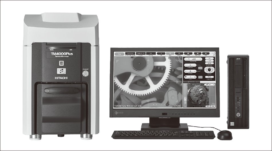
To support the study, advanced analytical instruments will be made available by Hitachi High-Tech IPC (Malaysia) Sdn. Bhd., including:
TM4000III Scanning Electron Microscope (lnkd.in/g3UMMjYV)
AZtecOne XploreCompact analytical system (lnkd.in/g8Q6VdME)
These tools will help researchers conduct detailed analysis, enabling both fundamental scientific research and applied investigations.
Beyond technology, the most meaningful outcome of this collaboration is the opportunity to build stronger academic partnerships in Malaysia and the broader ASEAN region. By working closely with researchers and institutions, we hope to contribute to the advancement of electron microscopy research while supporting the development of the regional scientific ecosystem. As research challenges become increasingly interdisciplinary, partnerships like these become essential to unlock new discoveries and applications.
Looking forward to seeing how this collaboration evolves in the coming years.
#ElectronMicroscope #Research Hitachi High-Tech Corporation
2
5
255

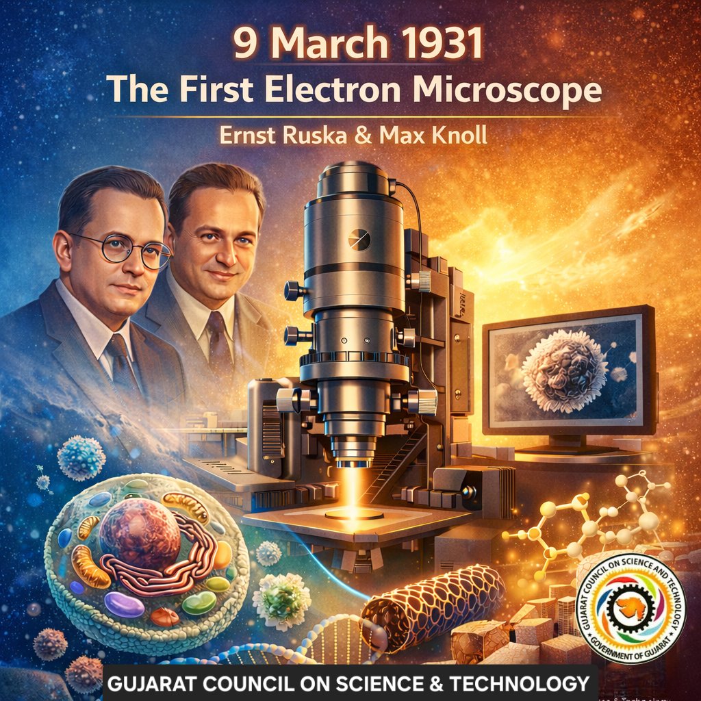
On 9 March 1931, science gained a powerful new vision into the unseen world!
German physicists #ErnstRuska and #MaxKnoll pioneered the invention of the first #ElectronMicroscope, opening a revolutionary window beyond the limits of #light microscopy and enabling #humanity to observe structures at an entirely new scale.
In recognition of his pioneering contribution to electron optics and the development of the first practical electron microscope in 1933, #ErnstRuska was awarded the #NobelPrize in #Physics in 1986.
From unveiling the intricate architecture of #cells and viruses to advancing the frontiers of #nanomaterials and modern #science, this landmark #innovation transformed #research across disciplines and continues to empower discoveries across #STEM, shaping our understanding of #life and matter at the smallest scales.
47
62
639
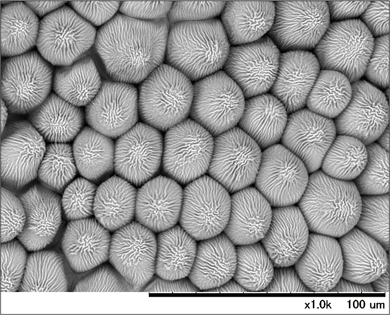
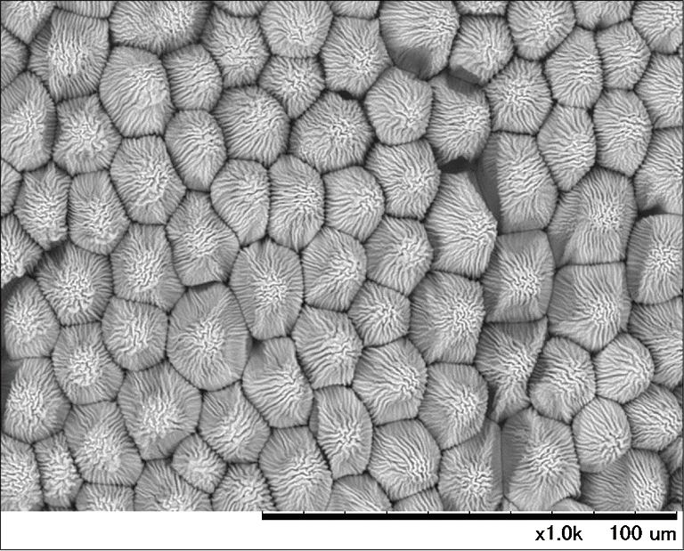
Happy Real Bread Week ! 🥖🍞🥐
(February 14–22, 2026)
#ElectronMicroscope #RealBreadWeek #BuddingYeast
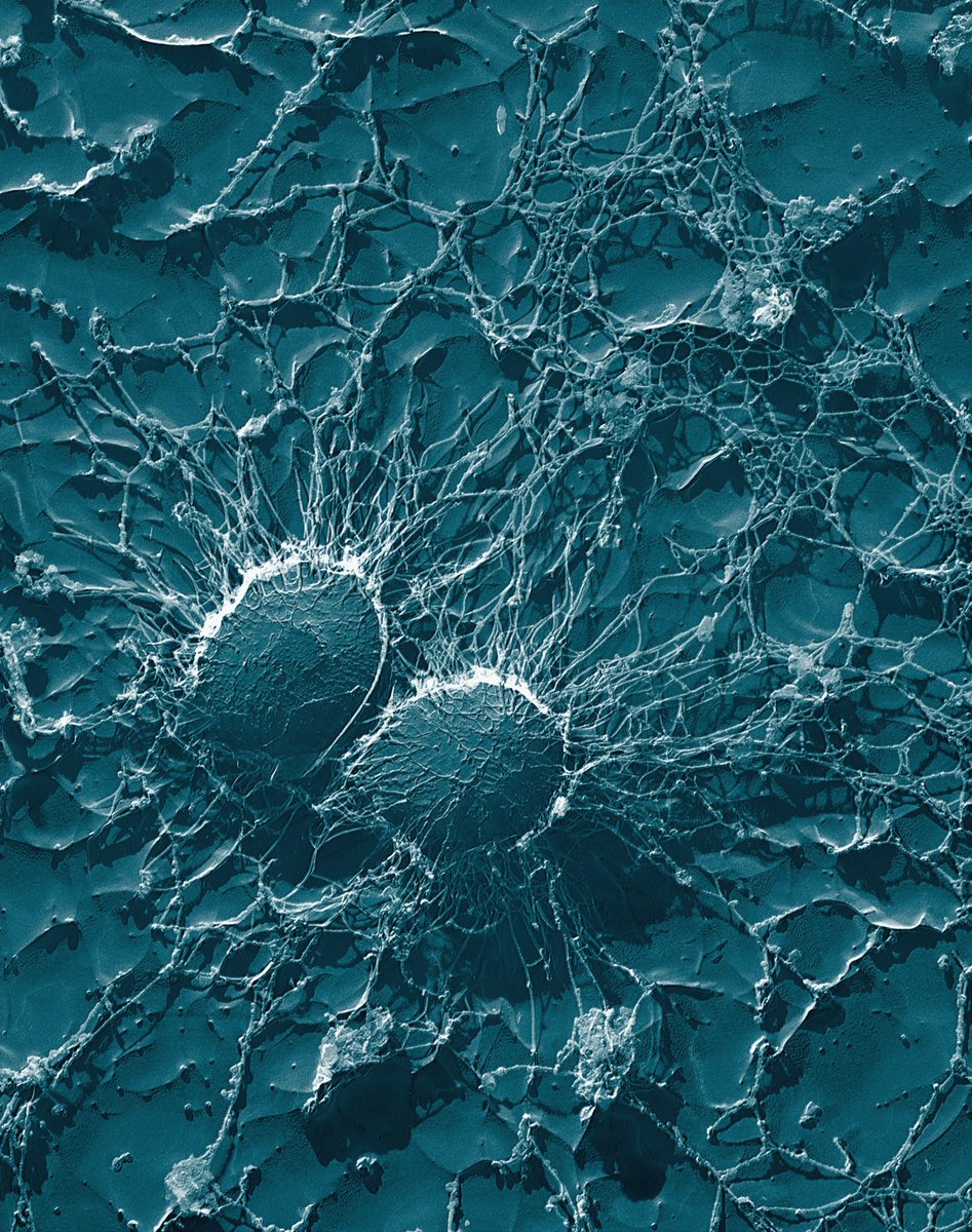
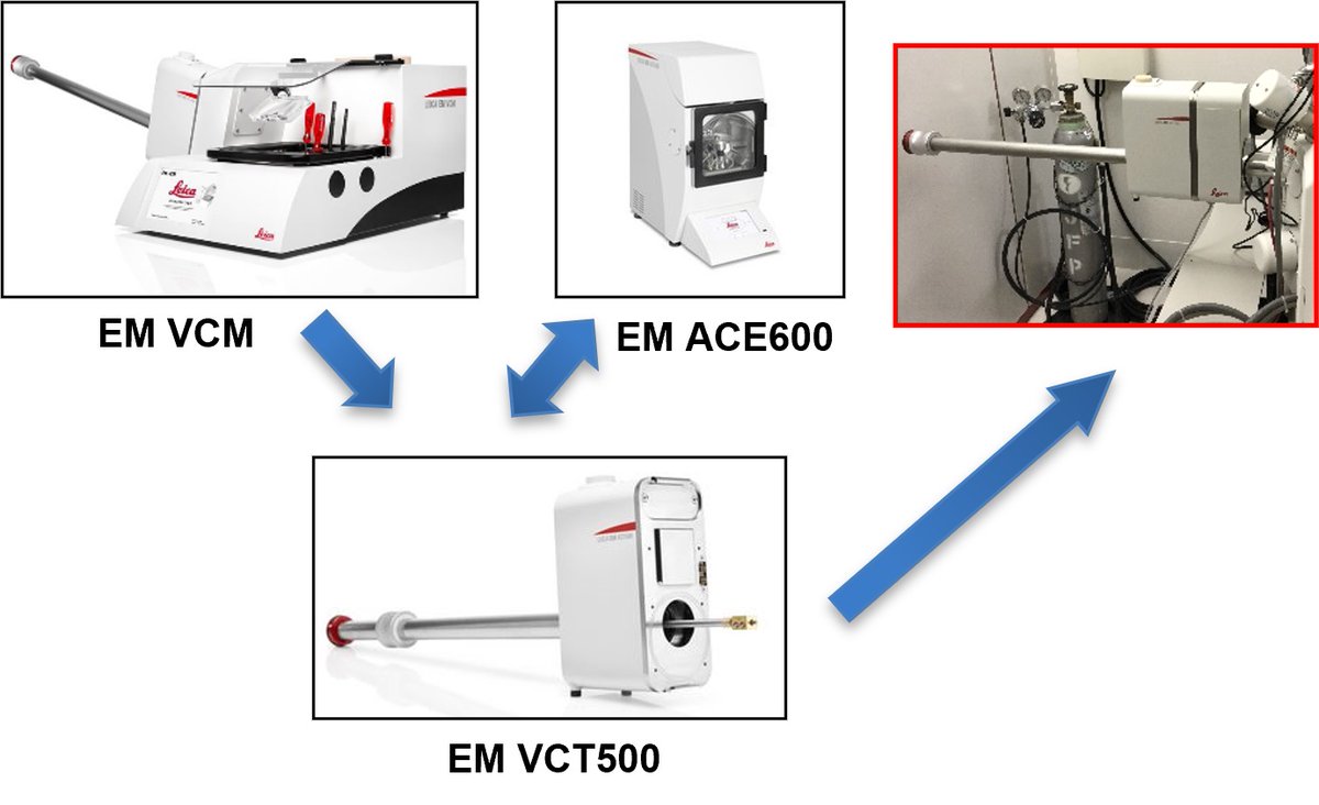
Budding yeasts were observed using an FE-SEM equipped with a cryo system. The yeasts were dropped directly onto a sample stub, rapidly frozen with slush nitrogen without prior fixation or dehydration, then cleaved and coated with 2 nm of Pt in the preparation chamber of the cryo system. Invagination structure unique to yeast cell membranes were observed in the exposed protoplasmic fracture (PF) face. In the enlarged view, particles with a diameter of approximately 15 nm were recognized and some were observed in hexagonal arrangement.
If you want to know more about the cryo imaging, check out the following post !
1
2
4
511

ಬರಿಗಣ್ಣಿಗೆ ಕಾಣದ ವಿಸ್ಮಯ: ವಿಶ್ವದ ಅತಿ ಸಣ್ಣ QR ಕೋಡ್ ಈಗ ಗಿನ್ನೆಸ್ ರೆಕಾರ್ಡ್ಗೆ ಎಂಟ್ರಿ! QR Code
vijayavani.net/category/news…
#ElectronMicroscope #WorldRecord #GuinnessBook #QRCode #TUWien
61
ಬರಿಗಣ್ಣಿಗೆ ಕಾಣದ ವಿಸ್ಮಯ: ವಿಶ್ವದ ಅತಿ ಸಣ್ಣ QR ಕೋಡ್ ಈಗ ಗಿನ್ನೆಸ್ ರೆಕಾರ್ಡ್ಗೆ ಎಂಟ್ರಿ! QR Code
vijayavani.net/category/news…
#ElectronMicroscope #WorldRecord #GuinnessBook #QRCode #TUWien
77
Happy Rose Day ! 🌹🌹🌹
(February 7)
#ElectronMicroscope #RoseDay #RosePetal
Electron Microscope images of a Rose Petal with and without cooling (at -20℃ vs RT)
This cooling stage allows samples to be cooled to temperatures as low as -25°C and maintained there for times ranging from tens of minutes to a few hours.
This reduces evaporation of water from moisture-containing samples, allowing observations and analysis to proceed without degradation of morphology.
The cooling stage is particularly well-suited to observations of samples with high water content—such as foodstuffs and biological tissues—or samples susceptible to thermal damage.
If you want to know more about the Tabletop Microscope with cooling system, check out the following post !
2
5
540
Happy National Beer Can Appreciation Day ! 🥫🍺🍻
(January 24)
#ElectronMicroscope #NationalBeerCanAppreciationDay #Aluminum
In-situ tensile testing of aluminum plate
If you want to know more about the testing, check out the following post !
3
578
Happy National Camcorder Day ! 📹🎥📷📱
(January 20)
#ElectronMicroscope #NationalCamcorderDay #AntiReflectionFilm
High resolution low voltage SEM of a moth-eye type anti reflection film
If you want to know more about the imaging, check out the following post !
1
2
409
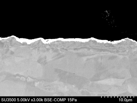
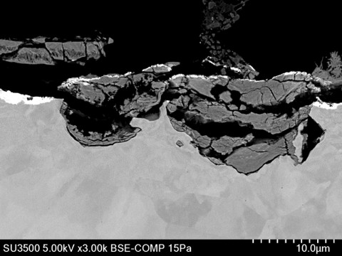
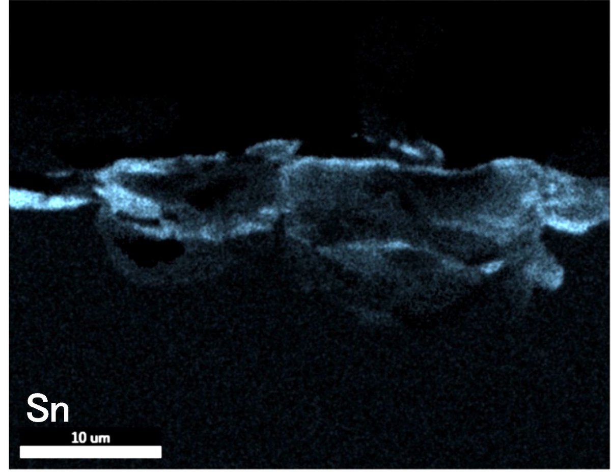
Happy National Tin Can Day ! 🥫🥫🥫
(January 19)
#ElectronMicroscope #NationalTinCanDay
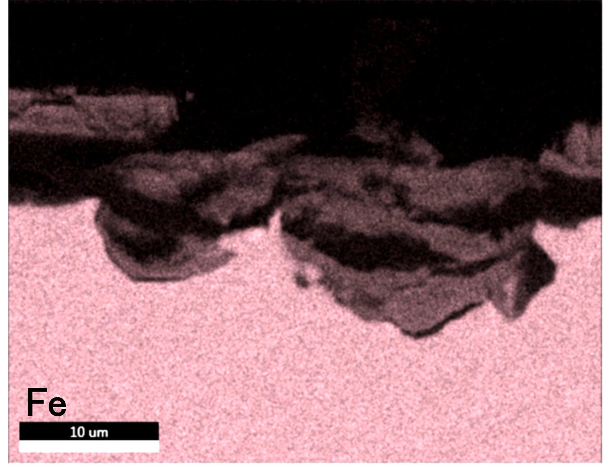
Normal and corroded areas of tin-plated steel sheet (Cross-sectional BSE images and elemental maps)
If you want to know more about the SEM and BIB (Broad Ion Beam) milling system, check out the following post !
3
8
879
Happy Book Publishers Day ! 📚📖📙🔖
(January 16)
#ElectronMicroscope #BookPublishersDay
SE image (surface information) and BSE image (compositional information) of a piece of Printed Paper
If you want to know more about the microscope, check out the following post !


3
367
Happy Argyle Day ! 🔶♦🔷
(January 8)
#ElectronMicroscope #electronmicroscopy #ArgyleDay
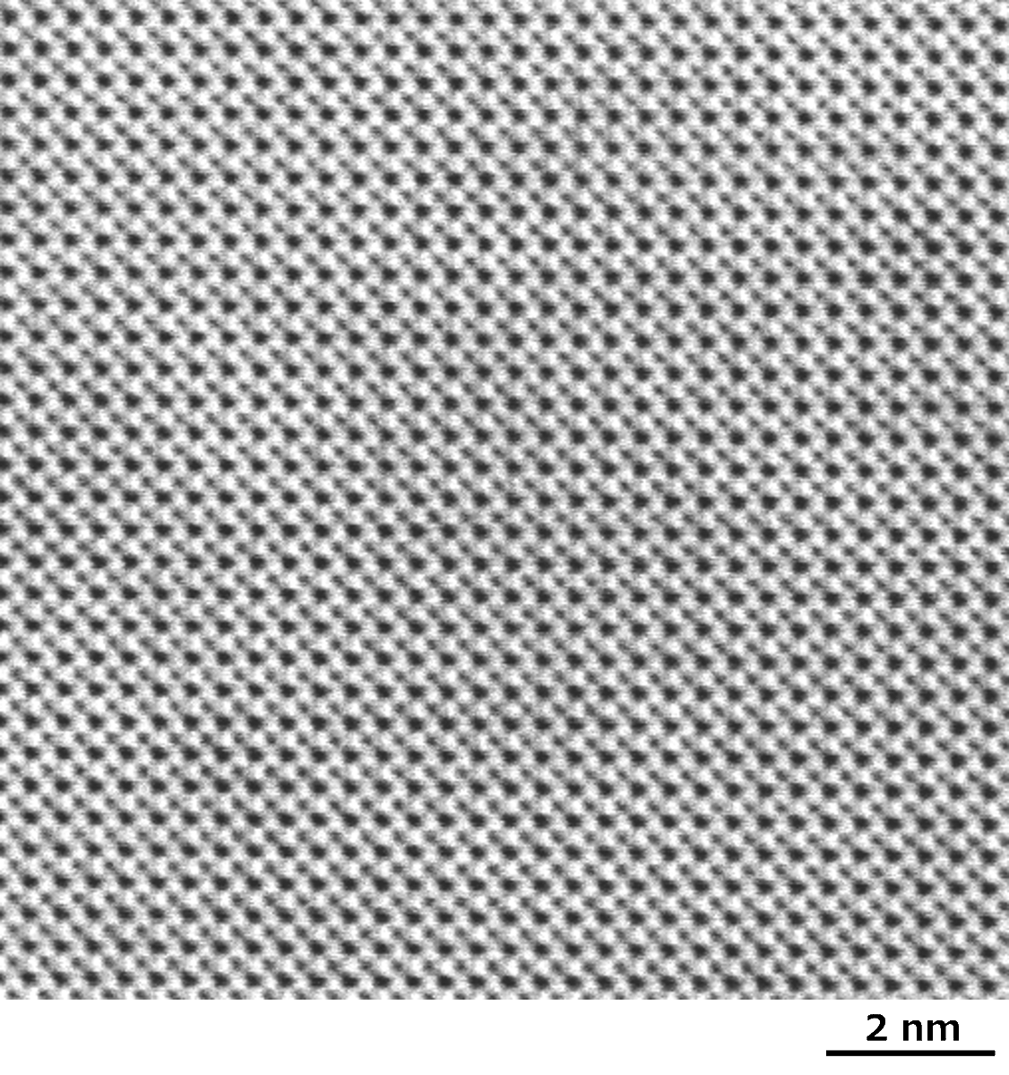
BF-STEM images of BaTiO3<001> single crystal
1
3
404

25 Dec 2025
Cross section of a TSV (Through Silicon Via) was prepared using BIB (Broad Ion Beam) milling system and the crystal orientation of Cu embedded in the TSV was analyzed by EBSD (electron backscatter diffraction). While the BSE image visualized each grain by channeling contrast, the EBSD map revealed that the grain sizes in the upper part are smaller than those in the lower part.
Even fine grains of several tens of nm were identified at higher magnification.
#TSV #ThroughSiliconVia #CrystalOrientation #BroadIonBeam #BIB #IonMilling #SEM #EBSD #ElectronMicroscopy #ElectronMicroscope
Available tools for this measurement are :
High Resolution Schottky Scanning Electron Microscope
hitachi-hightech.com/global/…
Further details and previously posted applications can be found on our YouTube channel. Looking forward to your visit.
youtube.com/channel/UC3zZlkQ…
3
617

8 Dec 2025
📺 “2009 abduction implant removal”
🔗 reddit.com/r/Interdimensiona…
“ For the first time, a doctor in the United States has come out with evidence that he claims is proof of alien visitations to Earth. This surgery has attracted world wide attention.
Who is Dr Roger Leir, DPM?
Dr Leir, DPM, is a surgeon from Southern California who has been performing operations on patients for over 30 years. He is a Podiatric Surgeon and has acted as Chief of Podiatry in many Southern California hospitals.
He lectures internationally on many medical subjects, and is a medical consultant to MUFON International. In the video he shows his most incredible and highly unusual finding, both biological and metallurgical, and presents the results from multiple independent scientific laboratories on the analysis of the implants!
Not just any lab, but Los Alamos National Laboratory, New Mexico Tech and the University of California, Santiago have analyzed this particular implant and have come to some astounding conclusions.
Amazingly, on analysis with a scanning electron microscope at NMT Laboratory, it was found that the implant WAS CONNECTED TO THE PATIENT’S NERVE ENDINGS! It was also covered by unidentifiable biological material that prevented ‘rejection’ by the patient’s immune system.
Implant connected to the patient’s nerve endings as seen through the electron microscope.
What’s more the lab tests conducted by NMT were corroborated by The University of California, Santiago which concluded that:
THE METALS AND THEIR COMBINATIONS IN THE IMPLANT WERE MANUFACTURED AND EXTRATERRESTRIAL IN ORIGIN.
Here’s the actual video of the implant being removed by Dr Roger Leir, DPM “
#AlienImplant #AbductionCase #AlienAbduction #ImplantRemoval #DrRogerLeir #UFOImplant #ExtraterrestrialEvidence #AlienSurgery #MUFON #LosAlamosLab #ElectronMicroscope #NerveConnection #MedicalAnomaly #IndependentAnalysis #MetallurgicalEvidence #UFO #UAP #Disclosure #AlienEvidence #CloseEncounters #ETContact #TruthIsOutThere #2009Case #UFOHistory #HiddenFiles #ClassifiedNoMore
1
2
174
24 Nov 2025
Happy National Cake Day ! 🎂🍰🍓
(November 26)
#ElectronMicroscope #electronmicroscopy
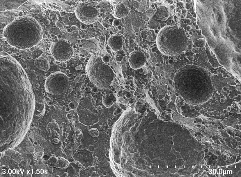
Cryo-SEM of air bubbles and fat globules in fresh cream
1
1
6
489
4 Nov 2025
It’s National Pathology Week !
🔬🧫🦠🧬💉
(November 3-9, 2025)
#ElectronMicroscope #electronmicroscopy #NationalPathologyWeek
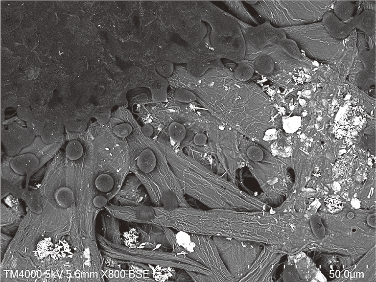
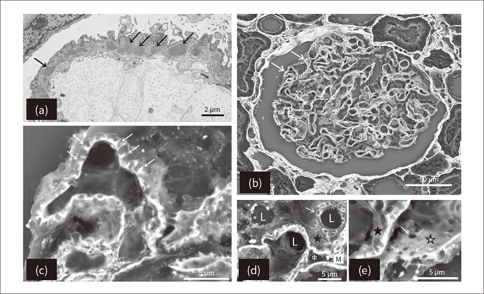
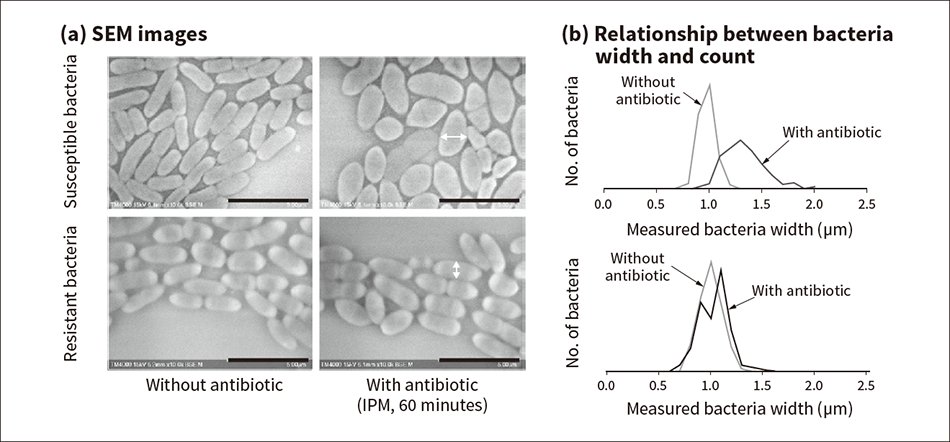
“Healthcare Applications for Tabletop Scanning Electron Microscopes
Microstructural Analysis beyond the Resolution of Optical Microscopes in Tissue and Bacterial Testing”
TEM and Low-vacuum SEM Images of Membranous Nephropathy of the Glomerulus
Read the full article here: hitachihyoron.com/rev/archiv…
1
5
620

31 Oct 2025
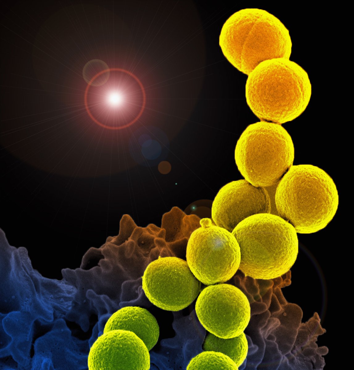
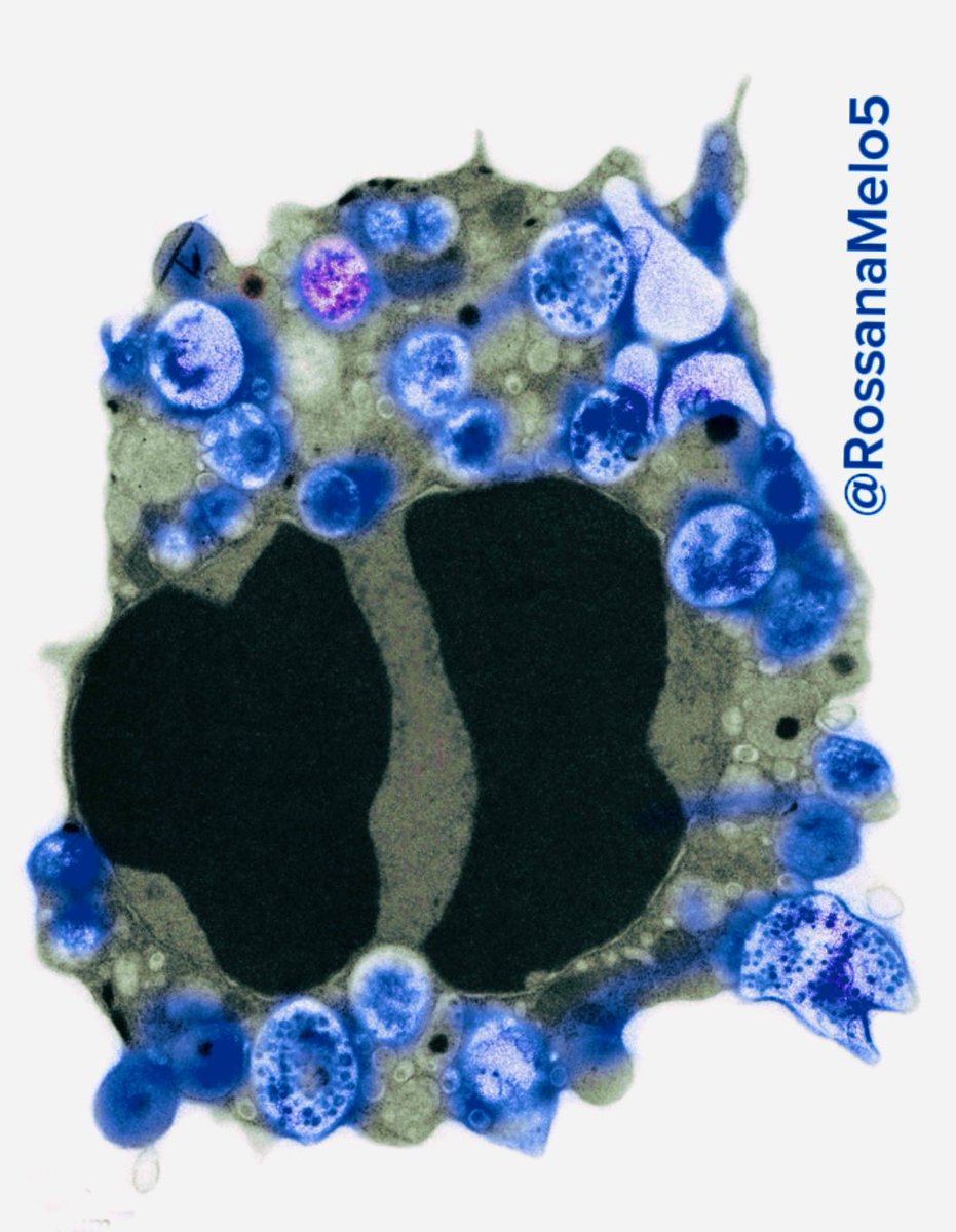
This #Halloween, meet the spookiest cell 👻: an #eosinophil undergoing #apoptosis, captured under the #electronmicroscope ! That highly condensed nucleus is so scary, it might haunt your dreams! 😱
2
8
272