
🔬 MetaboRamics: Raman imaging for live-cell spatial metabolomics, revealing dynamic metabolic rewiring in EMT
📊 Single-cell foundation models plateau at 1–10% of current datasets
🧭 CytoSignal: Infers ligand-receptor signaling at cellular resolution from spatial transcriptomics
1

🚨Lightning Talk alert at #ESHG!
👉Discover the power of genomics for new #RareDiseases diagnoses: “Multi-layer integration of long-read genome sequencing, transcriptomics and methylation outlier detection in 20 unsolved rare disease trios”
📌 Lightning Talks Stage 1
⏰ 13:10

14

Immunothrombosis in hospitalized COVID-19 patients identified by multiomics profiling and linked to postacute complications
🚨INTERESTING New Latvian/Swedish multi-omics study shows immunothrombosis never fully switched off in longC0VID patients: 3 months after COVID hospitalization, your blood is still biologically “clot-ready.”
➡️What makes this study so important:
1. It reinforces that immunothrombosis (the interplay of complement, NETs, and platelets) is a central driver in severe acute COVID-19 and can persist in longC0VID,
2. It confirms persistent endothelial Dysfunction/Endotheliopathy in longC0VID patients, consistent with earlier studies on vascular damage and microclots,
3. It aligns with prior evidence of mitochondrial dysfunction during the acute phase, followed by partial repair mechanisms,
4. The persistent complement activation they observed fits with other recent multi-omics studies showing ongoing complement dysregulation in longC0VID.
➡️Study:
1. Prospective longitudinal multi-omics study of 81 hospitalized COVID-19 patients tracked whole-blood transcriptomics, urine metabolomics (46 analytes), and 13 kidney-injury biomarkers at acute admission, ~1 month, and ~3 months post-hospitalization,
2. Patients stratified by EHR into recovered (n=35) versus long COVID (n=46) groups based on a PASC diagnoses within 12 months,
3. None of the 81 hospitalized patients were vaccinated,
4. Acute phase dominated by interconnected immunothrombosis: strong upregulation of complement (C1QA/B/C), NETosis (PADI4, MPO), and platelet-activation genes (ITGA2B, ITGB3), plus mitochondrial dysfunction (HIF1A/EPAS1 up, OXPHOS down, Warburg-like glycolysis) and elevated renal injury markers (KIM-1 etc.),
5. Most immune, mitochondrial, and metabolomic changes largely normalized by 1–3 months, with rebound in mitophagy/heme genes (PINK1, OPA1, FECH) indicating repair,
6. At 3 months, longC0VID patients showed a distinct transcriptional signature of persistent endothelial activation (↑VWF, PROS1, ITGA2B/ITGB3), complement dysregulation (CFH), and low-grade vascular inflammation/platelet reactivity (CXCL5, ALOX12) that was absent in recovered individuals,
7. No significant late differences in urine metabolomics or kidney biomarkers between groups.
➡️They conclude with their Highlight-points:
• Severe COVID-19 induces immunothrombosis-associated molecular programs,
• Acute COVID-19 is associated with mitochondrial metabolic dysregulation,
• Urine profiling indicates gradual renal recovery after hospitalization,
• LongC0VID patients retain endothelial-associated activation signatures.
‼️So, even after apparent clinical recovery, immunothrombosis leaves a persistent molecular scar of endothelial activation and prothrombotic signalling in longC0VID patients at three months, revealing that the acute vascular battlefield never fully quiets in those who remain symptomatic.
→Three months post Covid-19, longC0VID patient’s blood is still biologically primed to clot!
#AvoidSars2 #AvoidReinfections
cell.com/iscience/fulltext/S…

2
14
37
1,183
With OmicsLogic, you can explore Genomics, Transcriptomics, and Metagenomics through our Multi-Omics Course Track: omicslogic.com/programs/trac…

7

A fascinating new study reframes idiopathic inflammatory myopathies (IIMs): the key drivers of chronic muscle inflammation may not be immune cells alone, but tissue-resident fibro-adipogenic progenitors (FAPs).
Using single-nucleus RNA-seq, spatial transcriptomics, ATAC-seq, and primary human FAP cultures, researchers analyzed muscle biopsies from anti-synthetase syndrome (ASYS), inclusion body myositis (IBM), and immune-mediated necrotizing myopathy (IMNM). They found that FAPs adopt disease-specific inflammatory phenotypes that mirror the dominant immune environment.
Key findings:
🔹 IBM FAPs acquired T-cell–oriented programs, expressing mediators such as IL7 and CCL13.
🔹 IMNM FAPs preferentially engaged macrophage-associated inflammatory pathways.
🔹 ASYS FAPs displayed humoral immunity signatures and elevated IL6 expression.
🔹 Across all IIM subtypes, FAPs lost homeostatic markers (e.g., COL15A1) and shifted toward pro-inflammatory and pro-fibrotic states.
Trajectory analysis revealed two major FAP fates:
Homeostatic FAPs supporting muscle structure and regeneration.
Pro-inflammatory FAPs characterized by CXCL1, CCL2, IL18, LIF, COL1A1, COL1A2, LOX, and extracellular matrix remodeling programs.
Importantly, the proportion of pro-inflammatory FAPs increased with disease duration, suggesting progressive stromal reprogramming during chronic inflammation.
Mechanistically, the study identifies a dual-input signaling axis:
• Immune cells provide TGF-β signals.
• Damaged myofibers provide EGF signals.
Both converge on AP-1 transcription factor activity (JUN/FOS family), driving chromatin remodeling and establishment of a pathogenic FAP state. ATAC-seq demonstrated increased AP-1 accessibility after TGF-β EGF stimulation, while AP-1 inhibition reduced inflammatory cytokine induction.
Spatial transcriptomics further showed that FAPs form inflammatory niches by co-localizing with macrophages, muscle stem cells, and—in IBM—T cells, positioning them as organizers of local immune microenvironments rather than passive bystanders.
The broader implication is significant: chronic autoimmune muscle disease may involve a form of "stromal memory" or tissue priming, analogous to pathogenic fibroblasts in rheumatoid arthritis. If so, targeting FAP reprogramming, AP-1 signaling, BET proteins, or TGF-β pathways could complement conventional immunosuppression and potentially address treatment-refractory disease.
Reference
Nelke C et al. Inflammation reprograms fibro-adipogenic progenitors to sustain immunopathogenic niches in myositis. Cell Death & Disease (2026). DOI: 10.1038/s41419-026-08966-w.

69
Dr S S Hasan retweeted

Jun 5
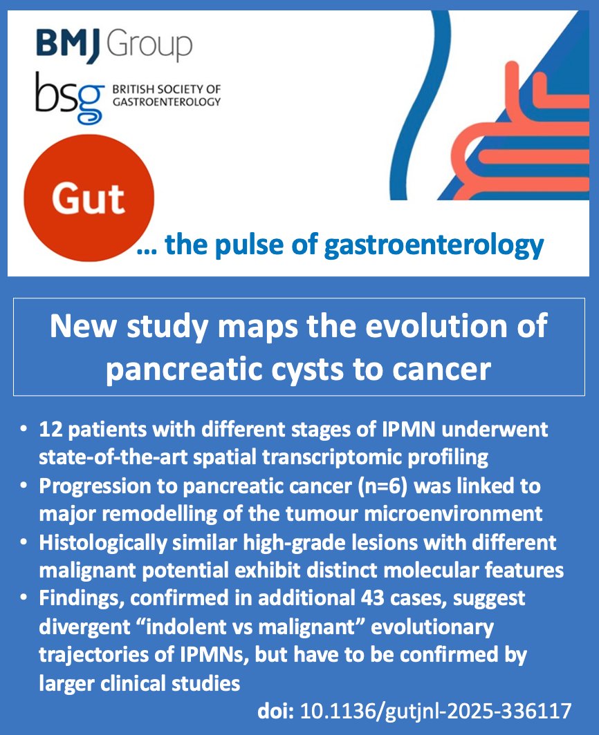
Important paper in Gut! 📢 New study maps the evolution of pancreatic cysts to cancer
Cui et al. used spatial transcriptomics to map progression from IPMNs to pancreatic ductal adenocarcinoma—revealing it’s not linear, but follows distinct molecular paths.
Early-stage lesions showed active immune surveillance (T cells and NK cells), while progression was marked by a shift to an immunosuppressive environment with expansion of regulatory T cells and fibroblasts.
Intriguingly, histologically similar high-grade lesions with different malignant potential were found to be molecularly distinct.
Malignant trajectories were characterised by upregulation of invasion-associated genes (e.g. CLDN1) TGF-β / IL-17 signalling.
⚖️ Are we over-treating some IPMNs while missing aggressive disease in others?
This study highlights the limitations of current risk stratification and the promise of spatially resolved biomarkers.
Check the paper: doi: bit.ly/4nIDprm
Social Media summary by Dr John P Thomas, Chain-Florey Clinical Research Fellow, Imperial College London, London, UK
3
6
606
Dr S S Hasan retweeted
Jun 12
Important paper in Gut! 📢 New study maps the evolution of pancreatic cysts to cancer
Cui et al. used spatial transcriptomics to map progression from IPMNs to pancreatic ductal adenocarcinoma—revealing it’s not linear, but follows distinct molecular paths.
Early-stage lesions showed active immune surveillance (T cells and NK cells), while progression was marked by a shift to an immunosuppressive environment with expansion of regulatory T cells and fibroblasts.
Intriguingly, histologically similar high-grade lesions with different malignant potential were found to be molecularly distinct.
Malignant trajectories were characterised by upregulation of invasion-associated genes (e.g. CLDN1) TGF-β / IL-17 signalling.
⚖️ Are we over-treating some IPMNs while missing aggressive disease in others?
This study highlights the limitations of current risk stratification and the promise of spatially resolved biomarkers.
Check the paper: doi: bit.ly/4nIDprm
Social Media summary by Dr John P Thomas, Chain-Florey Clinical Research Fellow, Imperial College London, London, UK

4
7
642

Cho et al. integrated whole-section (WS) spatial transcriptomics across 12 cancer types to construct a pan-cancer tertiary lymphoid structure (TLS) atlas. TLSs spanned early, primary, and secondary maturation states with distinct spatial niches and immune organization. Tumor regions proximal to intratumoral TLSs showed enriched antigen-presentation and IFN-response programs, and reduced proliferative and EMT signatures. An AI framework trained on whole-slide H&E images classified TLS maturation and maturation-aware TLS composite scores, which stratified survival and treatment response, outperforming conventional TLS metrics. bit.ly/4dZAbwi @mdAndersonNews @IamLinghua

5
23
1,133
antisense. retweeted

10 Jun 2025
PhenoGraph: A Multi-Agent Framework for Phenotype-driven Discovery in Spatial Transcriptomics Data Augmented with Knowledge Graphs biorxiv.org/content/10.1101/…

1
25
85
5,368
antisense. retweeted
7 Jun 2025
CellNEST reveals cell–cell relay networks using attention mechanisms on spatial transcriptomics nature.com/articles/s41592-0…

1
5
15
1,353

we worked on a transcriptomics project in cervical cancer & identified significant upregulation of glycolytic pathways.. highlighting the Warburg effect in tumor metabolism..
2
22

🧬 Gene expression profiling is indispensable for mapping cellular and molecular landscapes. Microarrays provide efficient, targeted analyses, while RNA-seq delivers deep, comprehensive transcriptomic insights. #GeneExpression #RNASeq #Microarrays #Transcriptomics #MolecularBiology #Genomics #PrecisionMedicine #Bioinformatics #CancerResearch #RareDiseases #NextGenSequencing #BiomedicalResearch #Omics #MolecularDiagnostics #LifeSciences #CRISPR #PGT #IVF #ART #NGS ⚕️

5

What’s New in Psoriatic Arthritis: A Whirlwind Tour
At #EULAR2026, Prof. Dennis McGonagle highlighted three key PsA trends: the rise of single-cell and spatial transcriptomics, progress toward personalized care (though not yet fully realized), and the next frontier of defining biologically refractory disease and exploring combination therapies.
buff.ly/pmHNYSn
5
4
473

Spatial transcriptomics revealed six serotonergic subpopulations in the dorsal raphe. Fluoxetine induced distinct acute and chronic gene-expression changes, affecting Htr1a, Ras/MAPK/cAMP signaling, axon guidance, and neuropeptides. nature.com/articles/s41380-0…
15

18h
CME INDIA Clinical Pearls
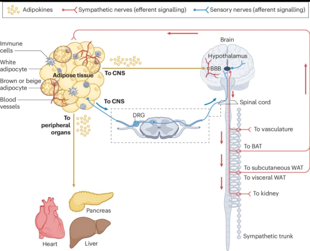
Adipose Tissue: The New Humoral–Neuronal Hub of Metabolic Regulation
(Nature Reviews Endocrinology, 2026)
🔹 1. Adipose tissue is no longer viewed as a passive fat storage depot. It functions as an active endocrine and neuroimmune organ that continuously communicates with the brain, liver, pancreas, heart, kidney, and skeletal muscle.
🔹 2. Adipose tissue communicates through two major pathways:
Humoral signals: adipokines, lipid mediators, metabolites, cytokines, and exosomal microRNAs.
Neuronal signals: sympathetic efferent and sensory afferent neural circuits.
🔹 3. The brain–fat axis is bidirectional. Adipose tissue sends metabolic information to the hypothalamus, while the CNS regulates lipolysis, thermogenesis, and energy expenditure through sympathetic outflow.
🔹 4. Sensory nerves within adipose tissue act as metabolic sensors. They detect thermal, mechanical, and chemical signals and relay this information to the CNS for rapid metabolic adaptation.
🔹 5. Sympathetic activation stimulates:
Lipolysis in white adipose tissue (WAT)
Thermogenesis in brown adipose tissue (BAT)
Browning of white adipose tissue
🔹 6. Cold exposure induces a unique endocrine program. Cold-activated adipokines and lipid mediators improve systemic glucose utilization, insulin sensitivity, and lipid metabolism.
🔹 7. Adipose tissue directly influences pancreatic β-cell function. Adipose-derived signals regulate insulin secretion and contribute to whole-body glucose homeostasis.
🔹 8. Extracellular vesicles and exosomal microRNAs are emerging as powerful endocrine messengers. These molecules enable adipose tissue to remotely modulate gene expression in distant organs.
🔹 9. Obesity causes "adipose communication failure." Both endocrine signaling and neural innervation become impaired, contributing to insulin resistance, chronic inflammation, and cardiometabolic disease.
🔹 10. Adipose neuropathy is an underrecognized consequence of obesity and diabetes. Loss of sympathetic and sensory nerve integrity disrupts lipolysis, thermogenesis, and metabolic flexibility.
🔹 11. Lipodystrophy represents the opposite extreme of adipose dysfunction. Deficient adipose tissue results in profound endocrine abnormalities and severe insulin resistance despite low fat mass.
🔹 12. Ageing is associated with progressive deterioration of adipose neuronal and humoral networks, contributing to frailty, sarcopenia, and metabolic decline.
🔹 13. The future of precision obesity medicine may involve targeting adipose communication pathways rather than simply reducing fat mass.
🔹 14. Emerging technologies transforming adipose biology include:
Single-cell multiomics
Spatial transcriptomics
Secretome labeling
Organoid models
Optogenetics
Click chemistry approaches
🔹 15. Therapeutic opportunities extend beyond GLP-1–based approaches. Synthetic adipokine analogues, lipid mediator mimetics, and neural circuit modulation may become next-generation metabolic therapies.
Take-Home Message
Adipose tissue is a sophisticated neuroendocrine organ that integrates humoral and neuronal signals to regulate whole-body metabolism. Obesity, diabetes, lipodystrophy, and ageing disrupt these communication networks, making adipose tissue signaling a promising frontier for future cardiometabolic therapeutics.
nature.com/articles/s41574-0…
4
9
428
ᗩᗷبᗩ𝕊 retweeted
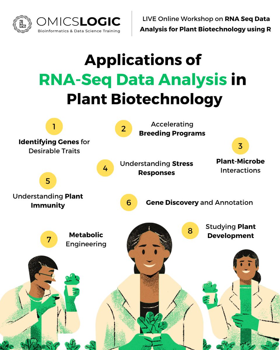
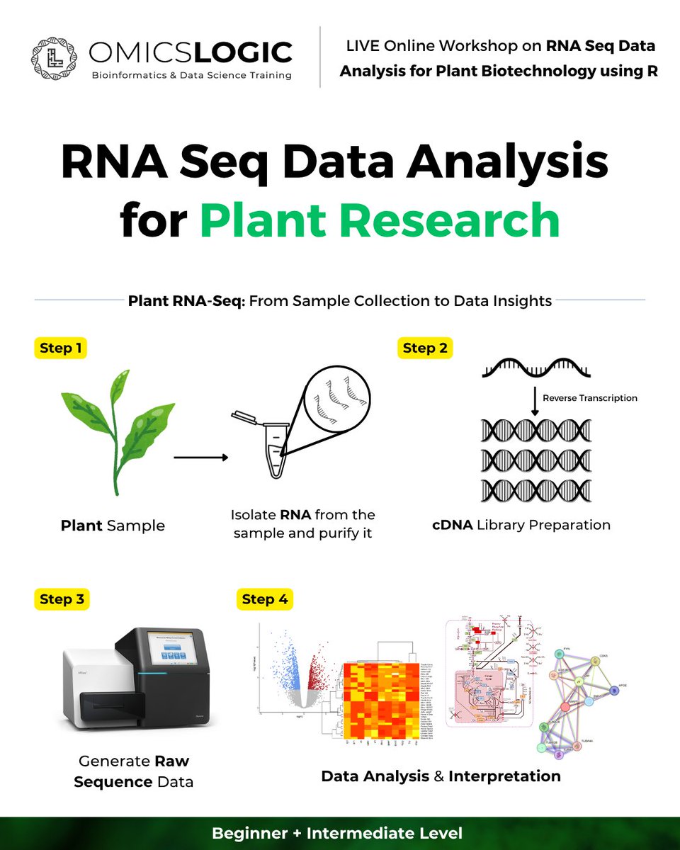
Unlock plant transcriptomics! 🌿 Join @OmicsLogic's 4-Day Workshop on Plant RNA-Seq Data Analysis with R. Learn to analyze nitrogen stress & tuberization workflows in potato. 🥔💻
🚀 Register: forms.gle/4bSsNJhZSLhVKsWG7
#Bioinformatics #RNASeq #PlantScience
1
1
22
ᗩᗷبᗩ𝕊 retweeted

Jun 12
🌱 Unlock plant transcriptomics! Join our 4-day live workshop on Plant RNA-Seq Data Analysis with R Programming to master end-to-end workflows for plant stress research.
🔗 Register: forms.gle/4bSsNJhZSLhVKsWG7
#Bioinformatics #RNASeq #PlantScience #RProgramming
1
2
35