
@UCBerkeley Metabolic Biology and Nutrition Twitter || Committed to #research, #education, and #innovation.
Joined December 2018
- Tweets 2,154
- Following 1,005
- Followers 1,213
- Likes 4,252
24 Photos and videos


UC Berkeley Metabolic Biology retweeted

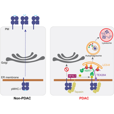
Our latest study out in @MolecularCell explores how and why MHC class I is aberrantly targeted for degradation via the autophagy-lysosome pathway in pancreatic cancer. Congrats to postdoc extraordinaire @marineberquez for leading this study! @UCSFAnatomy @UCSFCancer

May 29

Online Now: A multi-subunit autophagic capture complex facilitates degradation of ER-stalled MHC class I in pancreatic cancer dlvr.it/TSn7r3
2
21
149
30,957

UC Berkeley Metabolic Biology retweeted

May 21
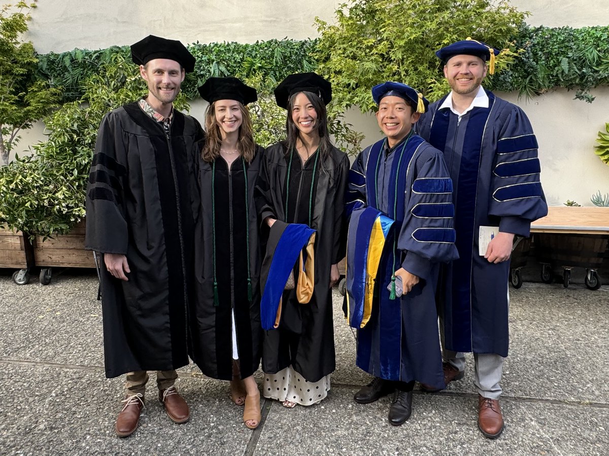
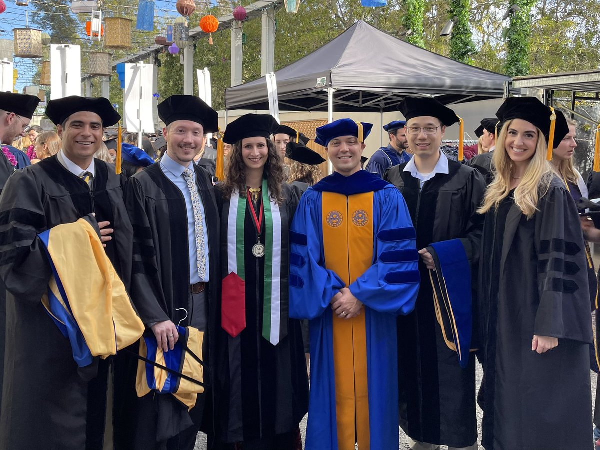
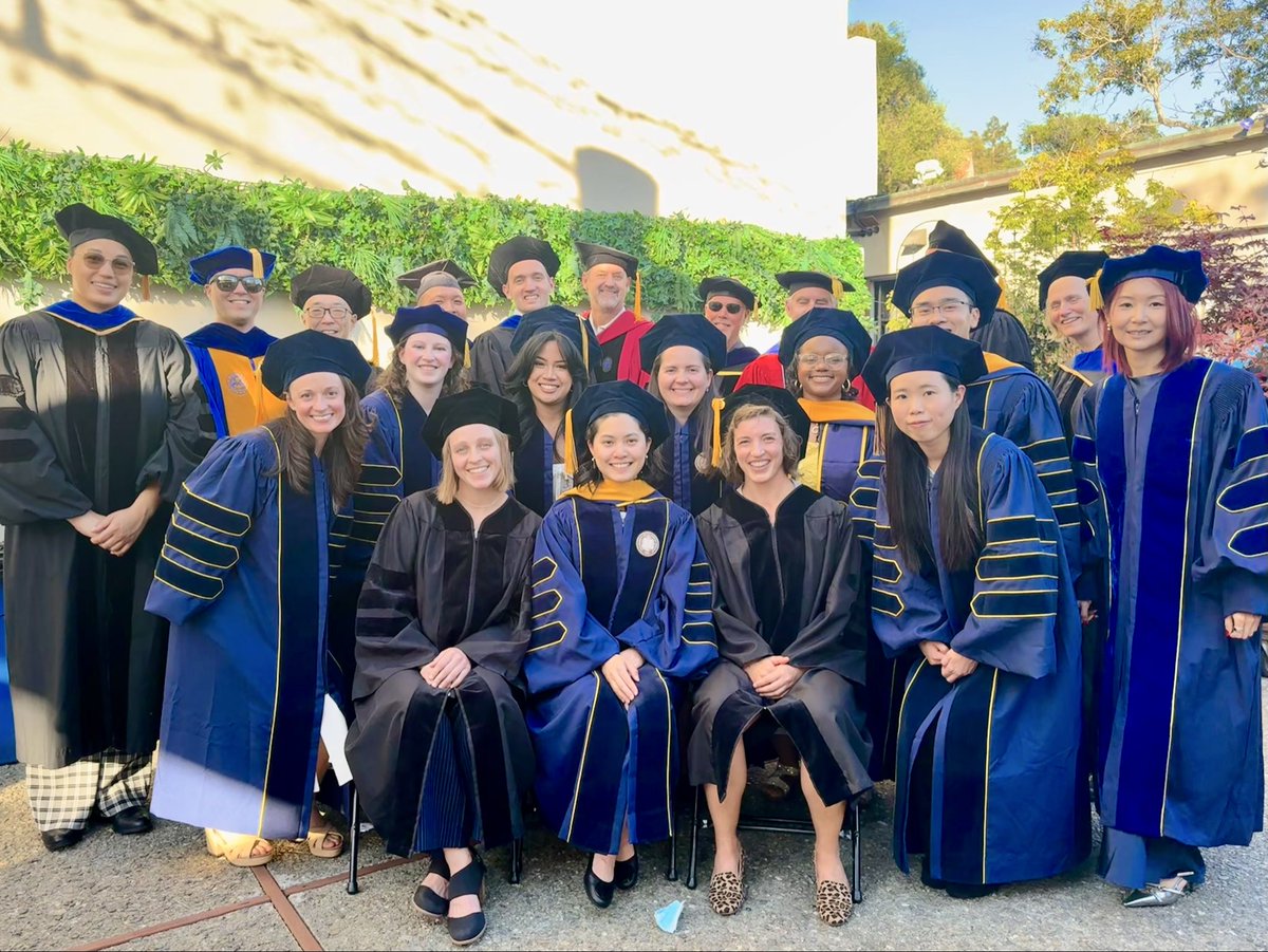
Congrats to chemical biology grad students Melissa Lim and Aman Modi from our lab for getting their PhD's!!

1
3
46
3,589
UC Berkeley Metabolic Biology retweeted
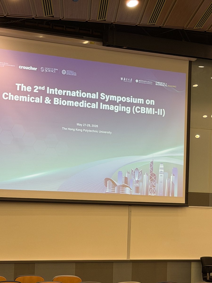
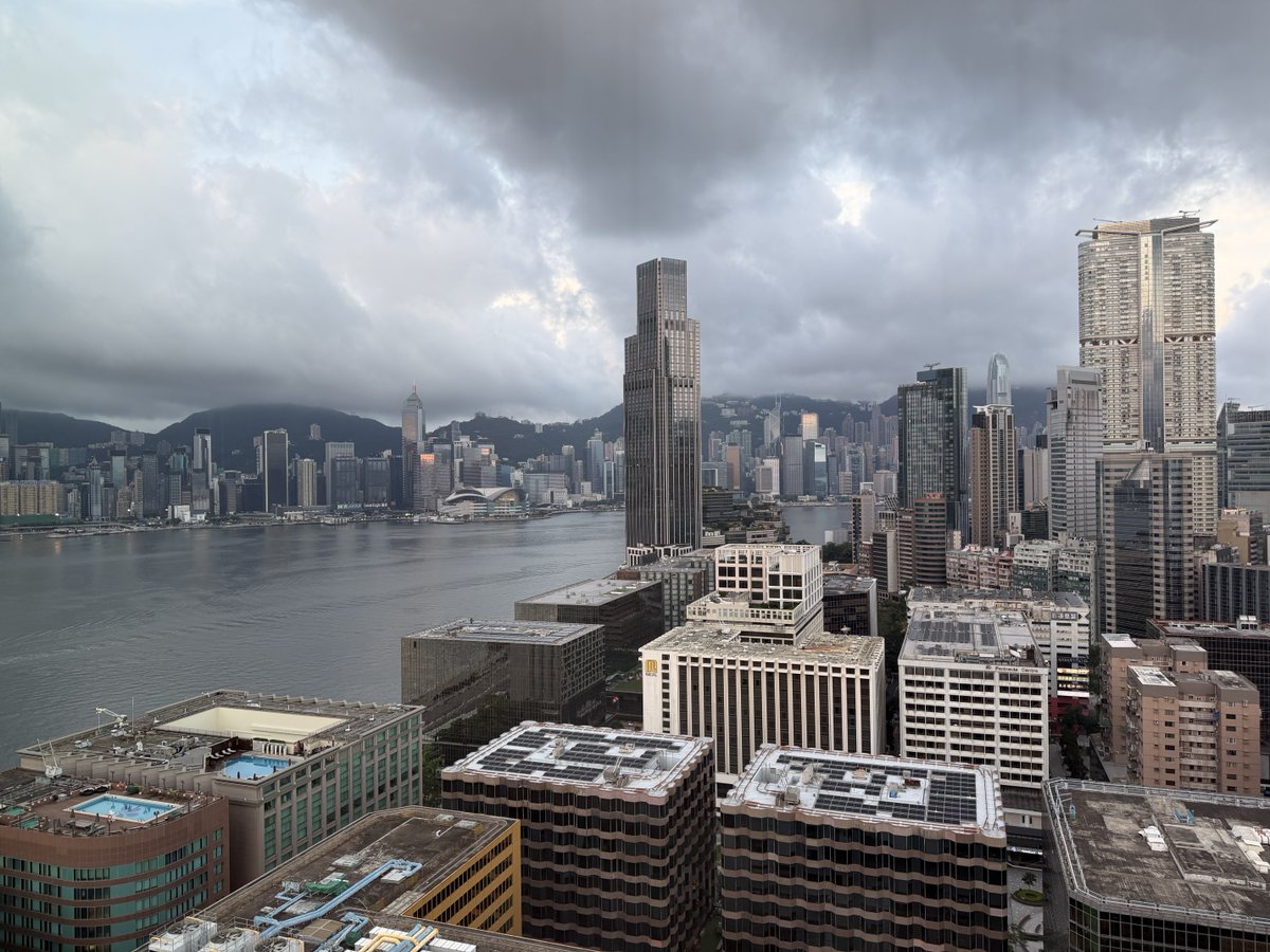
May 27
Excited to talk about our latest research at the 2nd International Symposium on Chemical and Biomedical Imaging in Hong Kong!
1
1
13
1,393

UC Berkeley Metabolic Biology retweeted

🌟 Welcome to Michael Ward, MD, PhD, joining @UCBerkeley as the inaugural Schekman Family Chancellor's Chair in Neuroscience, building on Berkeley's outstanding research in molecular and disease-related neuroscience.
➡️ bit.ly/4nBlOBo
@berkeleyMCB
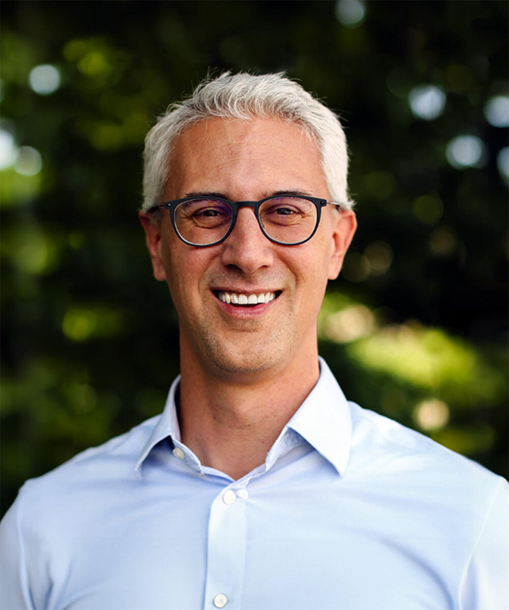
ALT Headshot of Michael Ward, a man with light hair, wearing glasses and a collared shirt
2
4
278
UC Berkeley Metabolic Biology retweeted

May 18
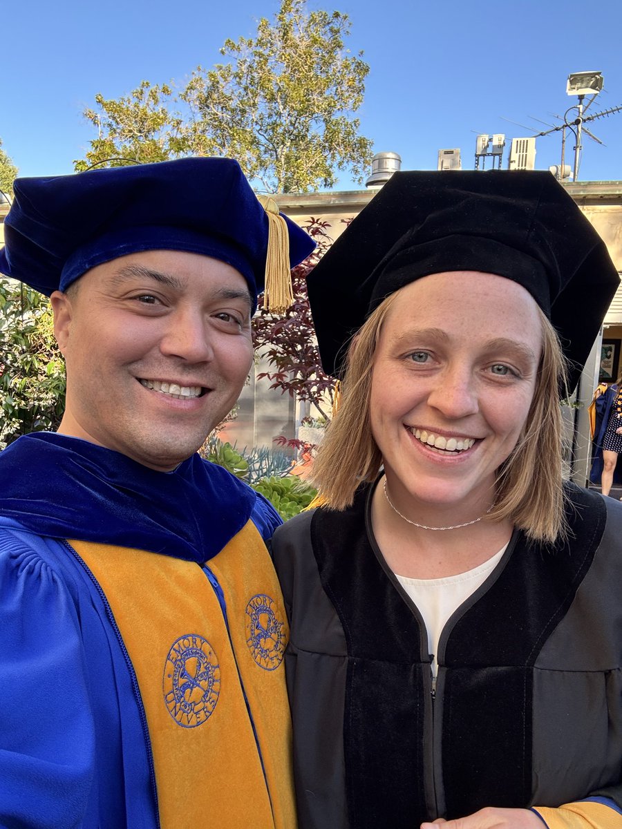
Proud to hood Sydney Tomlinson as she earned her PhD in Metabolic Biology at UC Berkeley. Sydney’s dissertation uncovered mechanisms for induced degradation of ER membrane proteins and advances our understanding of ER quality control.


1
3
56
3,342
UC Berkeley Metabolic Biology retweeted
May 19
Happy to have contributed to this preprint with Fangzhu Zhao and @realJimWells! LRP8-targeted bispecific antibody degraders degrade the selenium uptake receptor LRP8, reduce GPX4 / selenoproteins, and sensitize cancer cells to ferroptosis.
biorxiv.org/content/10.64898…

2
16
80
4,693
UC Berkeley Metabolic Biology retweeted
May 16
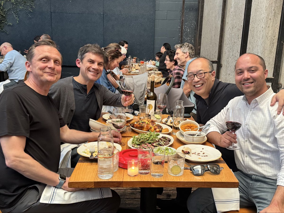
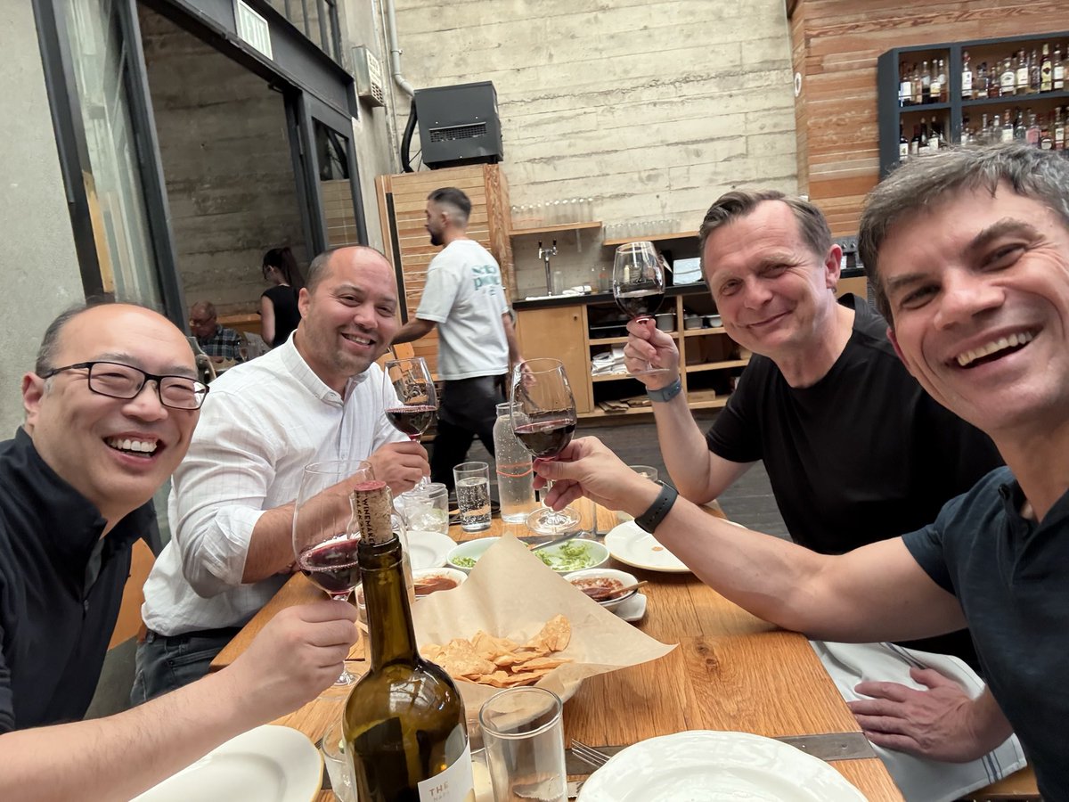
Some friendships just keep getting better with time. Really thankful for these guys, the conversations, the laughter, and the chance to slow down and enjoy an evening together. BFFs!☺️ @RobertoZoncu @RapeLab @DanNomura
1
3
54
4,255
UC Berkeley Metabolic Biology retweeted

May 7
Early iSOAR 5D movie of membrane and mitochondria dynamics over 3 hrs at 3 min intervals in the head of a zebrafish embryo 6 dpf, covering a 300 x 1000 um FOV at 193 x 193 x 550 nm resolution (0.8 TB total).
13 Dec 2025

Ever wonder why we decide to build a new microscope and how we go about it? For the last lecture this semester of the graduate microscopy class Na, Gokul, and I teach at Cal, I spoke on our developing iSOAR microscope, a swept HILO design with adaptive optics and structured illumination. iSOAR is designed to produce the petabytes of high resolution 5D videos of subcellular dynamics in zebrafish we need to train the multimodal vision models at the heart of our Cell Observatory Initiative. We still have some work to do, but the scopes are coming along nicely.
It's an exciting time at the Observatory, so if you're experienced and looking for a job in zebrafish transgenics, ultra-high throughput image processing, or the development of 5D spatiotemporal foundation models for interpreting the insane dynamic complexity of living matter, we'd love to hear from you. (sup@berkeley.edu, betzige@janelia.hhmi.org).
2
24
104
19,828

James Olzmann (@OlzmannLab at @UCBerkeley) won the 2026 Avanti Award in Lipids. His work has advanced knowledge of lipid droplets, lipid metabolism & ferroptosis.
⏯️Watch his #ASBMB26 talk, "Guardians of the membrane": youtu.be/QJ5YOsUCuHQ
#LipidResearch @AvantiResearch

ALT James Olzmann stands at a podium with a laptop delivering a talk at the 2026 ASBMB Annual Meeting.

8
41
3,754
UC Berkeley Metabolic Biology retweeted

Apr 17
Two historians, a biologist and a bioengineer from UC Berkeley have won illustrious Guggenheim Fellowships and will pursue independent work under “the freest possible conditions.” news.berkeley.edu/2026/04/17…
3
8
21
4,021
UC Berkeley Metabolic Biology retweeted

Apr 17
The first GLP-1 drug came from the Gila monster. But I think that is just one example of a much larger opportunity- harnessing natural products from extreme animals for human health. The feast-and-famine physiology of pythons is an ideal system to go fishing!! 🐍🐍🐍
6
11
92
11,117
UC Berkeley Metabolic Biology retweeted
Honored to recieve the 2026 UCSF Byers Award in Basic Science. Eternally grateful to my mentors, colleagues and friends for your support and to all the past and present Perera Lab members to whom this honor is dedicated to 🙏🏽


11
9
133
10,552
UC Berkeley Metabolic Biology retweeted

Apr 15
Congrats to MCB's Andreas Martin @AndyMartinLab on receiving the 2026 Dorothy Crowfoot Hodgkin Award from the Protein Society @ProteinSociety! 👏🎉
proteinsociety.org/protein-s…
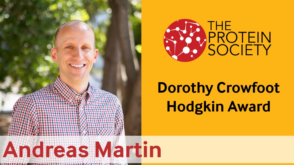
ALT Andreas Martin receives 2026 Protein Society Dorothy Crowfoot Hodgkin Award
1
4
315
UC Berkeley Metabolic Biology retweeted
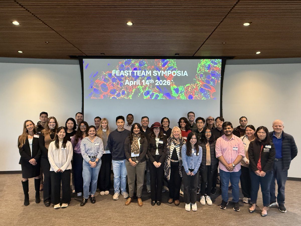
Apr 14
Team FEAST 🍔🍩🥑 of the Weill Cancer Hub West meets today @Stanford_ChEMH for our biannual symposium! With @rushika_perera @AttardiLaura @SuneilKoliwad @RuggeroDavide @RoarkeKamber @SvenssonLab @SCIDirector @StanfordCancer @UCSFCancer @Ashworth_SF
4
22
1,653
UC Berkeley Metabolic Biology retweeted

Apr 9
Interested in aging and rejuvenation?
We have a new Research Assistant position to understand aging at the cellular level!!
Come join our team!!! 😎
careersearch.stanford.edu/jo…
1
37
143
14,796
After spending much of his career building microscopes, work that earned him The @NobelPrize in Chemistry for contributions to super-resolution microscopy, Eric Betzig (@UCBIDS @hhmi_science) is turning his attention to the data those instruments produce.
youtu.be/FGZgAl1t9IU
3
4
452
UC Berkeley Metabolic Biology retweeted

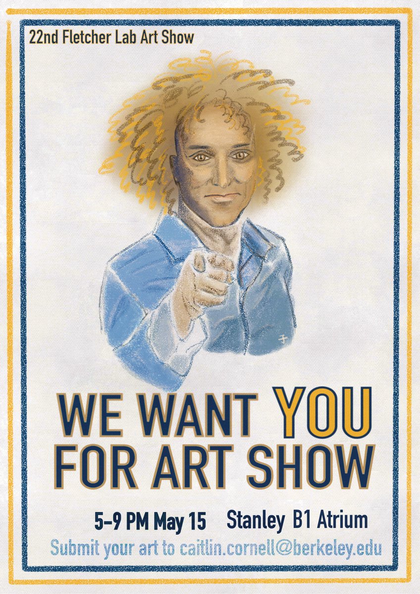
Apr 11
Join us for the 22nd Fletcher Lab Artshow! We’re looking for artists to submit their work and be part of an evening celebrating creativity and community. We will also have the return of the Ground State Ensemble! Art by @chunzi_liu
@QB3Berkeley
@UCB_Biophysics
@berkeleyMCB
4
9
1,083