
Jun 12
🔬 From advanced imaging technologies to training opportunities and community news, the latest @IctsRedib newsletter showcases how shared research infrastructures help drive biomedical discovery.
This issue highlights REDIB-2601, with 24 applications from 15 institutions, strong demand for expert support, and ReDIB’s continued move toward data-intensive biomedical imaging.
📝 Read the June issue: portal.redib.net/newsletters…
#CNIC #ICTSNews #ReDIB #BiomedicalResearch #ImagingScience | @GonzaloPiz_Card @balbella
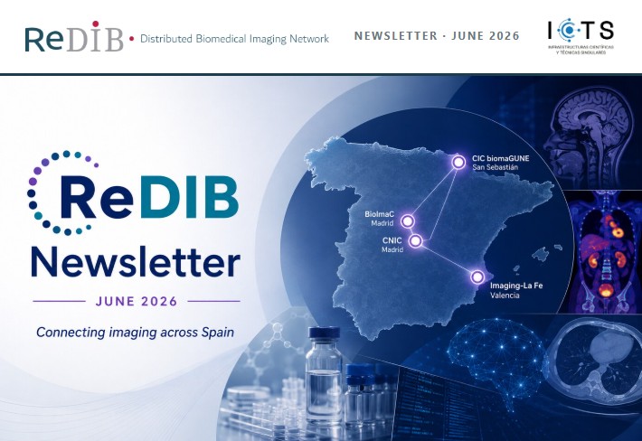
ALT ReDIB Newsletter June 26
115

May 2
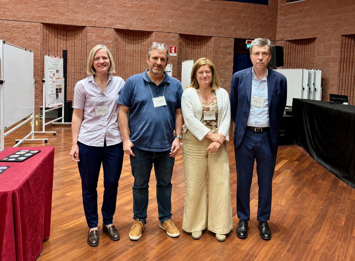
Glad that “Eye & Vision Session” was well received in @GordonConf on #ImagingScience . In 📸 Discussant Kristina Irsch (Institute de la Vision), Chair @sylvaingigan @Laboratoire_LKB , and speakers Susana Marcos @marcoslabur @cvsuor @UofR & @pablo_artal @Lo_um. Thanks all!!
1
1
4
328
May 1
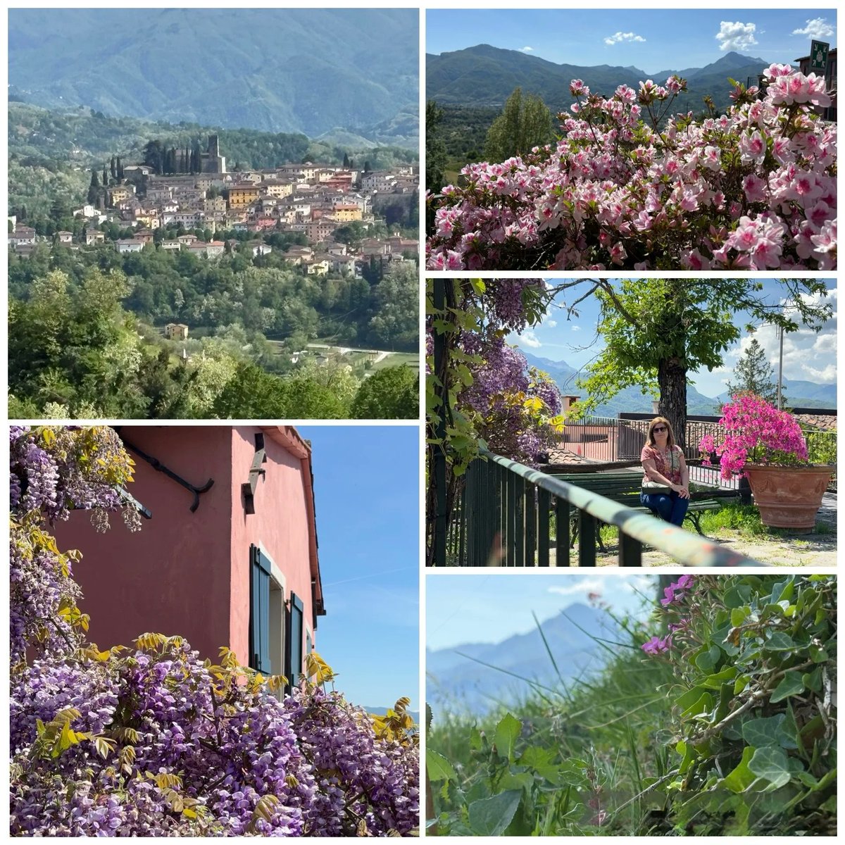
Attending the #GordonResearchConference on #ImagingScience in Tuscany. Wonderful location for meaningful scientific discussion without the rush of other conferences & to catch up cutting edge imaging technologies & applications. #GRC2026 @GordonConf
5
84

Mar 19
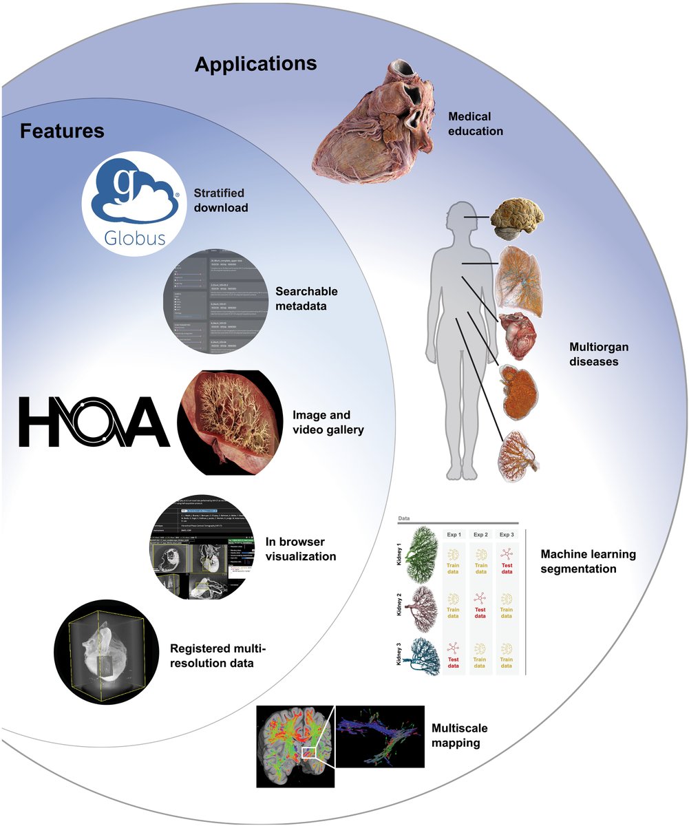
This is absolutely remarkable work from @ScienceMagazine. The Human Organ Atlas—built on Hierarchical Phase-Contrast Tomography (HiP-CT)—delivers something we’ve never had before: open-access, cellular-resolution 3D imaging of intact human organs, spanning healthy tissue and disease states. It’s a true public resource for researchers, clinicians, and educators.
The article is honest about the catch: HiP-CT needs a synchrotron (not exactly lab equipment). So while these atlases are phenomenal reference maps, their real power emerges only when any lab can bring their own data—microCT of a tissue block, light-sheet volumes, histology, whatever modality—and register it precisely against the atlas for direct, apples-to-apples comparison.
Next-gen imaging is now generating datasets at scales, resolutions, and multi-organ complexities that legacy analytical tools simply weren’t built to handle. The bottleneck has shifted from “how do we acquire the data” to “how do we make it interoperable, reproducible, and biologically meaningful.”
This is exactly we built the @neurosimplicity Imaging Suite: to close that integration gap with deterministic, multi-modal registration pipelines that let everyday labs leverage these gold-standard atlases without needing their own collider.
Huge respect to the HOA team—this is the kind of infrastructure that actually moves discovery forward.
Curious to hear from the community: how are you handling multi-scale data registration in your work today? What’s the biggest friction point you’re running into?
#HumanOrganAtlas #ImagingScience #OpenData #Neuroscience
Mar 18

The Human Organ Atlas, a new resource for researchers, clinicians, and educators, is an open-access database of 3D imaging of intact human organs.
The portal includes donor samples with conditions from congenital disorders to COVID-19.
Learn more in @ScienceAdvances: scim.ag/4bnSEzZ
3
18
3,832

Jan 20
Phase Retrieval: Recovering What You Can’t Measure
Lecture 1
One of the hardest inverse problems in science.
Do you know how your eye or a camera sensor actually sees an image? The sensor doesn’t record the wave itself. It records photon flux, which in wave language is intensity, a time-average of |field|². Phase still shapes what reaches the sensor through propagation and focusing, but at the pixel level the measurement is brightness.
So here’s the old, stubborn question behind a lot of optics, microscopy, astronomy, and wave imaging. If the object you care about is a complex field ψ(x), why does the world hand you only |ψ(x)|²? And when it does, what can you still recover about the phase that got erased at detection?
You have the field ψ(x) = r(x) exp(i θ(x))
Your detector stares at that and reports only
I(x) = |ψ(x)|² = r(x)².
The angle θ vanishes. So how do you get it back?
This lecture is the first, most basic approach one can think of...don’t try to make an intensity-only sensor magically phase-sensitive. Instead, force the phase to show up by interfering ψ with a known reference wave, and measure the intensity of the sum.
In the render, we do this with a normal image used as the amplitude r(x). The top-left panel is the only thing a sensor would give you directly...I(x) = |ψ|². The top-right panel is the interferogram Iᵤ(x;δ) = |ψ R e^{iδ}|² as a phase knob δ(t) is swept, so fringes slide even though r(x) stays fixed. The bottom-left panel isolates the signed cross-term (the phase leak) that drives those fringes. And the bottom-right panel is the payoff: after reconstructing θ̂(x) from four phase-shifted interferograms, we form ψ̂(x) = r(x) exp(i θ̂(x)) and display Re(ψ̂). That last image is not a phase color wheel...it’s a reconstructed wave-image built using the recovered phase.
The gentle math breakdown
Intensity hides phase for a single field, but it cannot hide phase for a superposition. Add a known reference wave
R(x) = A(x) exp(i φ(x))
and interfere it with ψ by forming
u(x) = ψ(x) R(x).
The detector measures
I_u(x) = |u(x)|² = |ψ(x) R(x)|².
Expand it
I_u
= (ψ R)(ψ* R*)
= |ψ|² |R|² ψ R* ψ* R
= I |R|² 2 Re(ψ R*).
That last term is where phase leaks back into something measurable.
Insert polar forms
ψ R*
= [r exp(iθ)] [A exp(−iφ)]
= r A exp(i(θ − φ)).
So the cross-term becomes
2 Re(ψ R*) = 2 r A cos(θ(x) − φ(x)),
and therefore
I_u(x) = r(x)² A(x)² 2 r(x) A(x) cos(θ(x) − φ(x)).
Now introduce the phase knob. Shift only the reference by a known δ
R(x) → R(x) exp(i δ).
Then φ(x) → φ(x) δ, so
I_u(x; δ) = r² A² 2 r A cos(θ − φ − δ).
So a single intensity image deletes θ, but a controlled family of interferograms forces θ to show itself through a predictable cosine swing.
#PhaseRetrieval #Interference #Optics #ImagingScience #Holography #ComputationalImaging #SignalProcessing #Mathematics #Physics
5
11
67
4,036

Jan 19
How fast do proteins travel along a neuron’s axon? 🧠
Now, with MI-SIM super-resolution imaging, we can directly measure their speed and track dynamic docking events in living cells!
📹 Study from Prof. Li’s lab at Tsinghua University
#ImagingScience #Neuroscience
5
190

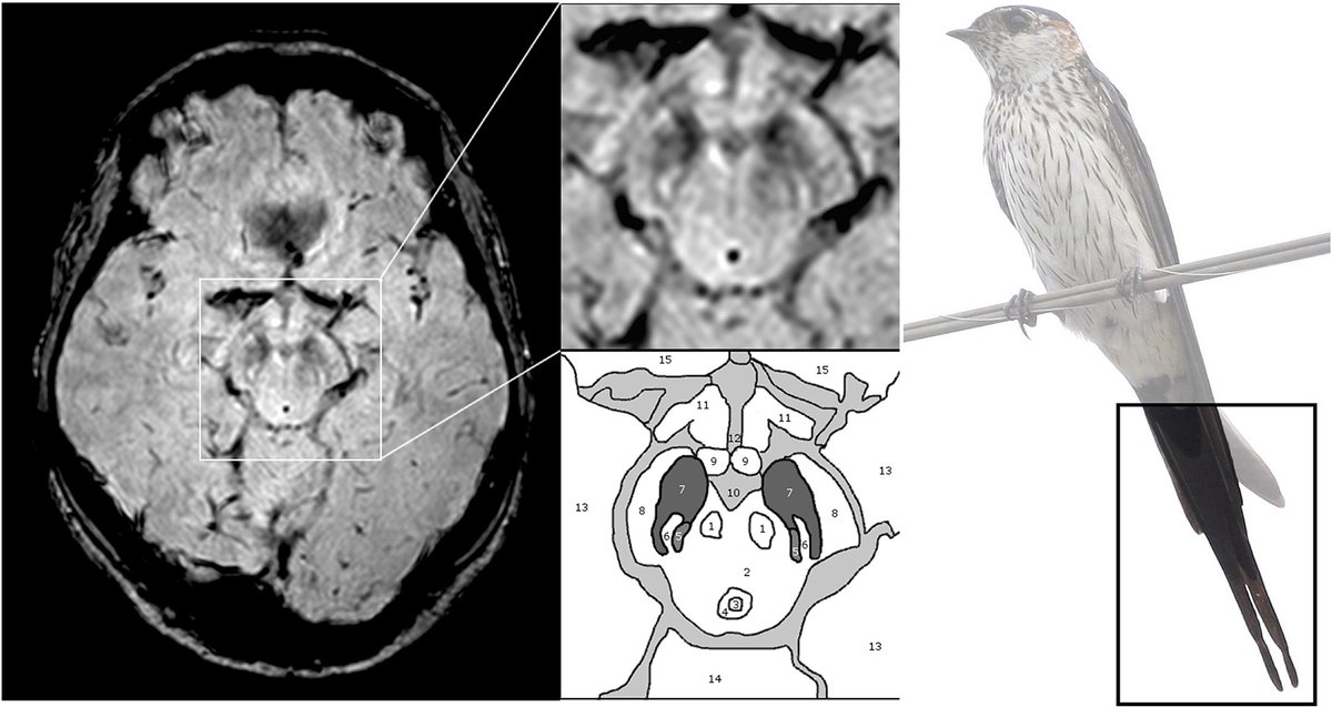
Parkinson’s disease can be hard to distinguish from its mimics. A recent Radiology review highlights MRI biomarkers like Nigrosome 1 and neuromelanin that, combined with standard imaging, improve diagnostic confidence and guide dopamine transporter imaging.
Read the full article: bit.ly/48y9qwl
#NeuroRad #ImagingScience
3
28
163
7,274

21 Nov 2025
📣🎉 We’re delighted to share that IC Dean and director of Imaging and Visual Representation Laboratory, @ssusstrunk, has received the 2025 @The_RPS award for #ImagingScience.
👏 Warmest congratulations to Professor Süsstrunk on this remarkable honor!
actu.epfl.ch/news/sabine-sus…
8
284

17 Nov 2025
It was terrific having our entire software development team at @circleoptics HQ at @nextcorps last week. You can hear from each of the team members in our software development series on Stitchless_Circle Optics in focus :youtube.com/playlist?list=PL… #TeamCircle #CircleOptics #imagingscience #softwaredevelopment 1 @AnjaliJogeshwar
1
2
61

3 Nov 2025
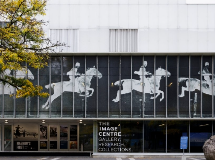
I see every day, walking to my office, the iconic frames of "The Horse in Motion" @TorontoMet 's The Image Centre: capturing movement once invisible to the human eye (reminded also of the bet businessman Leland Stanford wanted to settle! )
Today, we use high-speed microbubble "photography" in the lab to reveal bubble motions that are too fast to see to better understand how microbubbles respond to #ultrasound
More on Eadweard Muybridge’s "Animal Locomotion" & how his work contributed to modern cinema 👇
torontomu.ca/news-events/new…
#ImagingScience #Ultrasound #Microbubble #Science
1
2
204

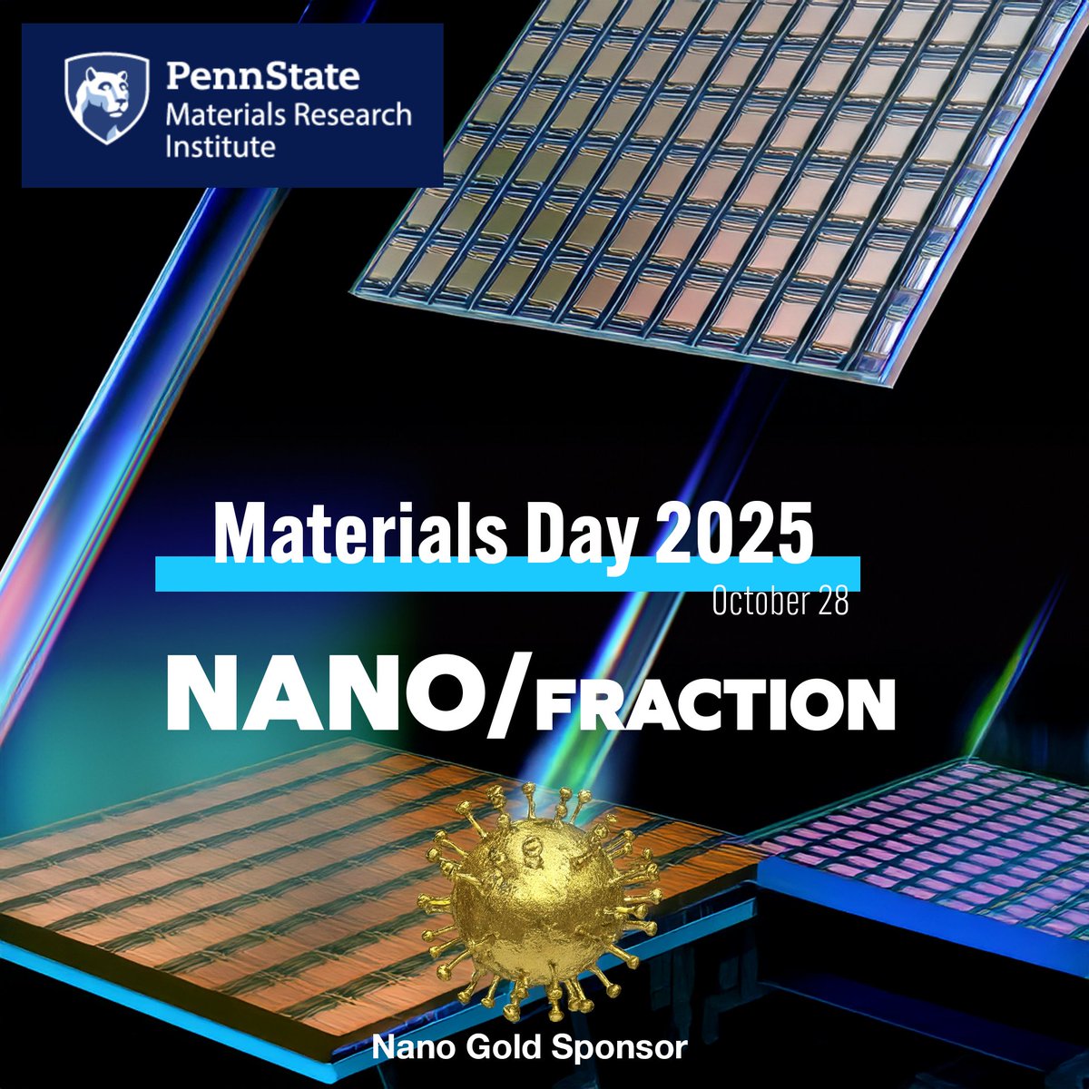
NanoFraction is proud to sponsor this year's Material Day 2025. Come visit us on October 28th at the Materials Research Institute to learn more about Optical Super Resolution. mri.psu.edu/events/2025-mate…
#Superresolution #Nanotech #PennStateResearch #DrugDelivery
#Microfluidics #Innovation #NanoFraction #MaterialsDay2025 #PennStateMRI #PennState #StateCollege #OpticalSuperResolution #NanoscaleImaging #MaterialsScience #Semiconductors #Photonics #Microscopy #LabelFreeImaging #Metrology #DefectDetection #WaferInspection #QuantumMaterials #ThinFilms #Research #RAndD #STEM #DeepTech #LabToFab #Innovation #HardwareStartups #ImagingScience #PrecisionManufacturing #TechEvent #Sponsor
1
2
129

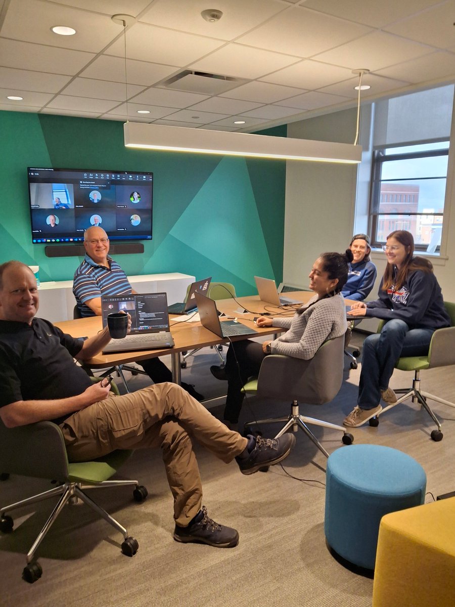
Cherry Huang @cherryhuang0120 presenting at @DnbResearch‘s seminar with the kind of slides that stops you mid-note: rigor wrapped in beauty, intricate image analysis unraveling the #cerebellum’s secret migration choreography and . #Neuron #ImagingScience #Science #cellbio

8
767

Interested in #ImagingScience in #PaediatricOncology? 🔍
Join CCLG’s first-ever Imaging Science Day in person on Friday, 5 September. Discover exciting new developments and research from expert speakers.
🚨 Registration deadline: 22 August
Register at bit.ly/45cyibe
2
3
237

🔥 NEW SESSION ALERT!
Hot Topics in Bioimage Analysis & AI is coming to #SPAOM 2025! 🤖🔬
Cutting-edge tools, deep learning, explainable AI & more — all in one session. 💥
Don’t miss this! 🚀
#AI #BioimageAnalysis #Microscopy #DeepLearning #ImagingScience #HotTopics
6
12
801
Interested in #ImagingScience in #PaediatricOncology? 🔍👨🔬
Join CCLG’s first-ever Imaging Science Day in person on Friday, 5 September. Discover exciting new developments and research from expert speakers.
More info and register at bit.ly/45cyibe

4
247

8 Aug 2025
📢 New Publication in @JACCJournals : Cardiovascular Imaging
“Staging of Cardiac Adverse Remodeling in Moderate or Severe Aortic Regurgitation” by @MaanMalahfji and colleagues.
In this prospective study, the authors investigate a proposed cardiac remodeling staging system for patients with moderate or severe aortic regurgitation. Such a staging system that helps define the extent of cardiac remodeling can provide important prognostic value in AR beyond traditional markers, particularly when guideline thresholds are not met.
🫀 As clinicians move toward earlier diagnosis and management of AR, this work supports the role of advanced cardiac imaging in shaping clinical decision-making.
Read the full open access article here: sciencedirect.com/science/ar…
@PhilGenereuxMD @SachinGoelMD @DipanJShah
#Cardiology #WhyCMR #AorticRegurgitation #JACC #StructuralHeart #HeartValveDisease #CMR #ImagingScience #JACCImaging #ValveTherapy

5 Aug 2025

We report a staging system of Cardiac Adverse Remodeling in Chronic AR, analogous to the AS system by @PhilGenereuxMD | JACC: Cardiovascular Imaging @HMethodistCV @DipanJShah @WilliamZoghbi @SachinGoelMD
jacc.org/doi/10.1016/j.jcmg.…
3
3
7
1,732

31 Jul 2025
We’re honored to share some exciting news!
The USC Brain Tumor Center has been selected as a 2025 Wright Foundation Transformative Cancer Grant Award recipient for our proposal:
“Multi-parametric Dynamic Contrast Imaging (mp-DYCI) of Glioblastoma for Early Prediction of 6-Month Outcomes and Accurate Diagnosis of True Progression.”
This groundbreaking project aims to advance how we assess and treat glioblastoma by developing more precise imaging tools to differentiate true tumor progression from treatment effects—earlier and more accurately than ever before.
We’re incredibly grateful to the Wright Foundation for supporting this innovative research, and proud of our team for continuing to push the boundaries of what’s possible in brain cancer care.
Stay tuned as we move this work forward—and thank you to all who make this progress possible.
#CancerResearch #Glioblastoma #NeuroOncology #USCBTC #MedicalInnovation #ImagingScience #WrightFoundation

4
8
358

31 Jul 2025
🎓 PhD Opportunity at Vrije Universiteit Amsterdam (VU Amsterdam) , Netherlands | PhD position in Ultra-High Speed Computational Optical Coherence Tomography (OCT)
📲 phdscanner.com/phd-vacancies…
📍 Amsterdam, Netherlands
📅 Application Deadline: October 31, 2025
📧 Contact: Dr. Dierck Hillmann (d.w.a.hillmann@vu.nl)
We're hiring! The Faculty of Science at VU Amsterdam is offering an exciting PhD position in Ultra-High Speed Computational Optical Coherence Tomography (OCT). Join a cutting-edge research team exploring the intersection of optics, computational imaging, and biomedical innovation.
✔️ Develop experimental setups for full-field Fourier-domain OCT
✔️ Advance image reconstruction algorithms using sparse sampling and compressive sensing
✔️ Collaborate closely with ARCNL
✔️ Contribute to high-impact publications and conferences
✔️ Thrive in a dynamic, diverse, and interdisciplinary research environment
💼 Your profile:
✔️ MSc in Physics, Engineering, or related field
✔️ Strong background in optics/imaging
✔️ Skilled in Python, C/C , or MATLAB
✔️ Curious, creative, and collaborative mindset
💰 Salary: €2,901 – €3,707/month (gross)
⏳ 4-year contract with full academic and career development support
🌍 Located at the vibrant and accessible Zuidas campus in Amsterdam
🔗 Apply by October 31, 2025 – early applications encouraged!
📄 Submit: Cover letter (max 1 page), CV, recent grades, and (if possible) a written report in English.
Let's push the boundaries of biomedical imaging—together!
#PhDposition #OpticalImaging #OCT #ComputationalImaging #BiomedicalResearch #VUAmsterdam #PhDNetherlands #ScienceJobs #AcademicJobs #ImagingScience
1
2
215

4 Jul 2025
🧠🔬 📸High schoolers explored the power of scientific imaging with Daniel Sage during the pre-university weeks organized by the Education and Science Outreach department at @EPFL_en
#FutureScientists #ImagingScience #EPFL

5
213

2 Jul 2025
🔬 3D volume scans are essential in neuroscience!
While a single soma 🧠 might look crisp in a 2D plane, its dendrites are another story — branching out in every direction like neurons dancing under the microscope 🌐✨ (not the sun... unless your microscope is really bright).
Capturing the full picture means thinking in volumes, not just slices.
#Neuroscience #Microscopy #ImagingScience #BrainResearch #Neuroanatomy #Dendrites #3DImaging #VolumeScanning #Neurotech #LifeInTheLab
1
2
120