
22 Dec 2025
📿 Medical Pearl
Category: USMLE Step 1 > Pathology > Cardiovascular Pathology
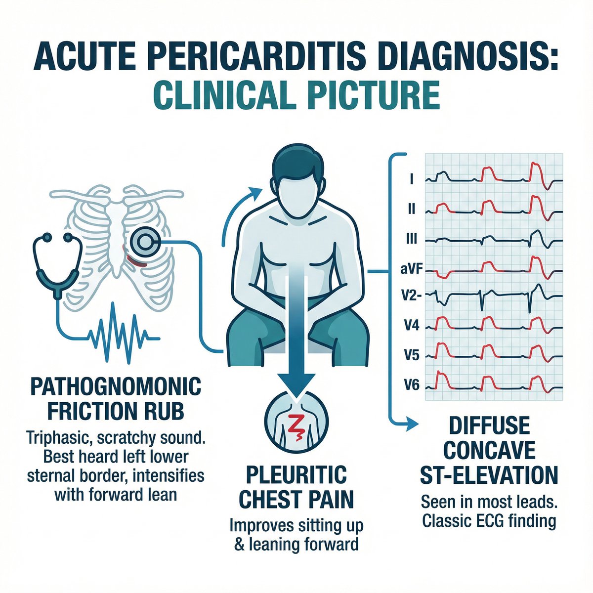
Acute pericarditis diagnosis is a clinical picture. Suspect it with pleuritic chest pain that improves sitting up and leaning forward. Auscultate carefully for the pathognomonic, scratchy, triphasic pericardial friction rub, often best heard along the left lower sternal border, intensifying with forward lean. Crucially, the ECG typically shows diffuse, concave ST-segment elevation in most leads, accompanied by PR-segment depression, particularly with reciprocal PR elevation in aVR. This specific ECG pattern, in conjunction with the pain and rub, strongly differentiates pericarditis from ischemic cardiac events. Echocardiography helps assess for effusion or tamponade, but the bedside clinical and ECG findings are usually diagnostic.
🎬 Watch the video explanation: youtube.com/watch?v=36ki7njD…
🔗 Learn more: endlessmedical.academy/auth?…
#MedStudent #MedStudentTwitter #ClinicalPearl #FOAMed #MedEd #Medicine #ClinicalMedicine
@MedTwitJC @MedEdChat @MedEdPortal @MedTwitter @MedPedsJournal @bmaboreddr @MedStudentTwit
Dig deeper on this topic at endlessmedical.ai, where multiple AI models debate, cross-check references, and verify calculations to give you the most updated and accurate medical information with minimal hallucinations and errors.
References:
1. 2025 ESC Guidelines for the management of myocarditis and pericarditis (Inflammatory Myocardial and Pericardial Syndromes) (2025) (escardio.org/Guidelines/Clin…)
2. A Randomized Trial of Colchicine for Acute Pericarditis (ICAP) (2013) (pubmed.ncbi.nlm.nih.gov/2399…)
3. Pericarditis — NHS (2023) (nhs.uk/conditions/pericardit…)
*Generated by AI. May contain errors. Use at own risk. Full disclaimer: endlessmedical.academy/auth?…
2
38
22 Dec 2025
📿 Medical Pearl
Category: USMLE Step 1 > Pathology > Neuropathology
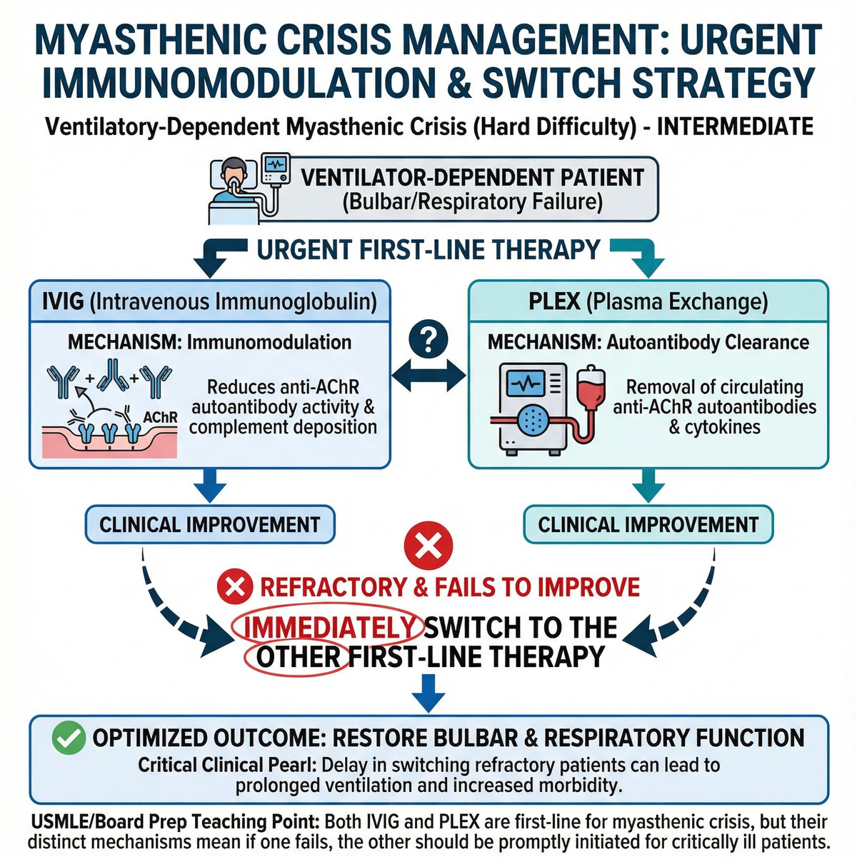
Myasthenic crisis demands urgent, effective immunomodulatory therapy. While intravenous immunoglobulin (IVIG) and plasma exchange (PLEX) are both first-line options, their mechanisms differ. For critically ill patients in a ventilatory-dependent myasthenic crisis, if one modality proves refractory and fails to improve bulbar or respiratory function, immediately switch to the other first-line therapy. Do not re-dose the ineffective treatment. This strategy ensures rapid removal of pathogenic autoantibodies or modulation of the immune response through a different pathway, crucial for preventing prolonged ventilation and improving clinical outcomes. Promptly escalating to the alternative is key.
🎬 Watch the video explanation: youtube.com/watch?v=U29tl3dr…
🔗 Learn more: endlessmedical.academy/auth?…
#MedicalEducation #MedTwitter #ClinicalMedicine
@NEJM @MedEdChat
Dig deeper on this topic at endlessmedical.ai, where multiple AI models debate, cross-check references, and verify calculations to give you the most updated and accurate medical information with minimal hallucinations and errors.
References:
1. International Consensus Guidance for Management of Myasthenia Gravis: 2020 Update (2021) (pmc.ncbi.nlm.nih.gov/article…)
2. Intravenous immunoglobulin for myasthenia gravis (2021) (cochrane.org/evidence/CD0022…)
3. FDA approves inebilizumab-cdon (Uplizna) for generalized myasthenia gravis (2025) (mda.org/press-releases/fda-a…)
4. Myasthenia Gravis – NINDS Fact Sheet (2020) (ninds.nih.gov/publications/m…)
*Generated by AI. May contain errors. Use at own risk. Full disclaimer: endlessmedical.academy/auth?…
2
59

17 Oct 2025
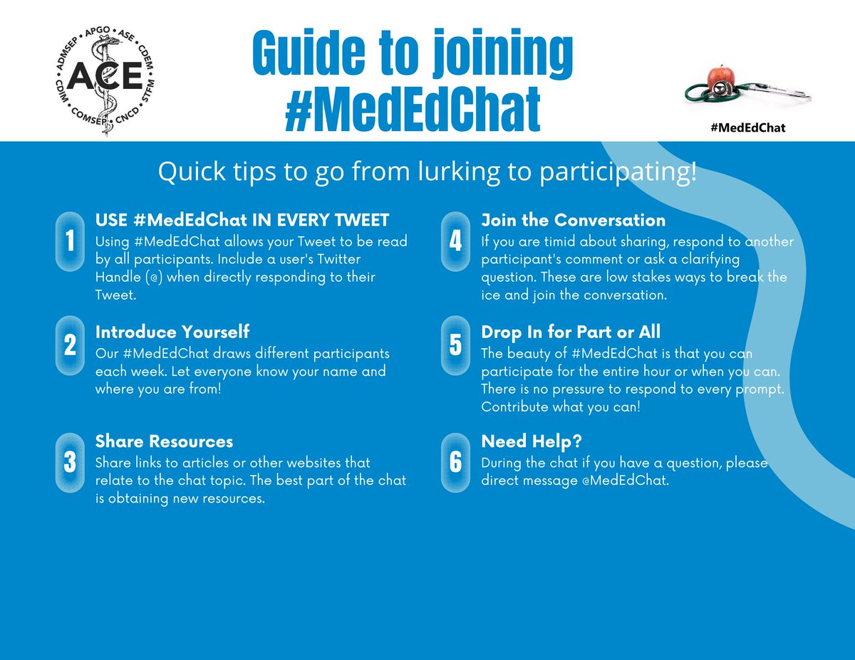
New #MedEdChat blog!
More med students. Fewer clinical teachers. Our latest post explores how to motivate & support physician faculty who make #Meded possible. EVUs, tax incentives, and smarter systems—what really works?
allianceforclinicaleducation… @admsep @Alliance4ClinEd
2
143

16 Jul 2025
This pattern (leg >> arm weakness) points to an Anterior Cerebral Artery (ACA) stroke. Clinical pearl: The subtlest finding—like pronator drift—is often the key to localization. #NeuroTwitter #FOAMed @AANMember @MedEdChat
1
1
3
178

14 May 2025
A.I. and Remote Learning: The End of the Academic Model for Medical Schools?
indd.adobe.com/view/426718bb…
@MedEdChat @DrQuinnCapers4 @IhabFathiSulima

1
2
899


25 Jan 2025
“It is not enough to have a good mind; the main thing is to use it well”
—RENE DESCARTES
#MedTwitter #Motivation #UsmleCoach #MedEdChat
1
590

5 Nov 2024
Happy November!! From >450 med ed pubs in Oct, we picked 3 MUST READS. Content areas: bedside teaching, partnering with pts, & 'time' in med ed. @MedTeachJournal @asmeofficial @ameefacdev @AAMCtoday @acgme @GIMMedEdDoc @CLOSLER @MedEdChat @MedEdPORTAL
hopkinsbayviewinternalmedici…
6
5
963
9 Sep 2024
Final third of the year! Time flies. To help you out, we've selected 3 MUST READS from >450 published med ed articles last month; they touch on clin reasoning, unions, & assessments. @MedTeachJournal @AcadMedJournal @mededice @AAMCtoday @ameefacdev @acgme @GIMMedEdDoc @MedEdChat
2
4
910

27 Jun 2024
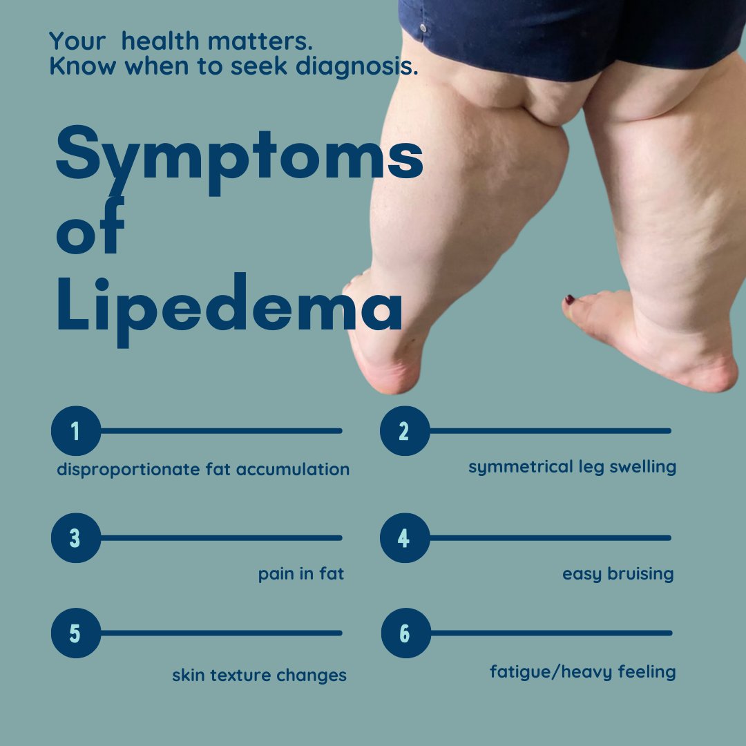
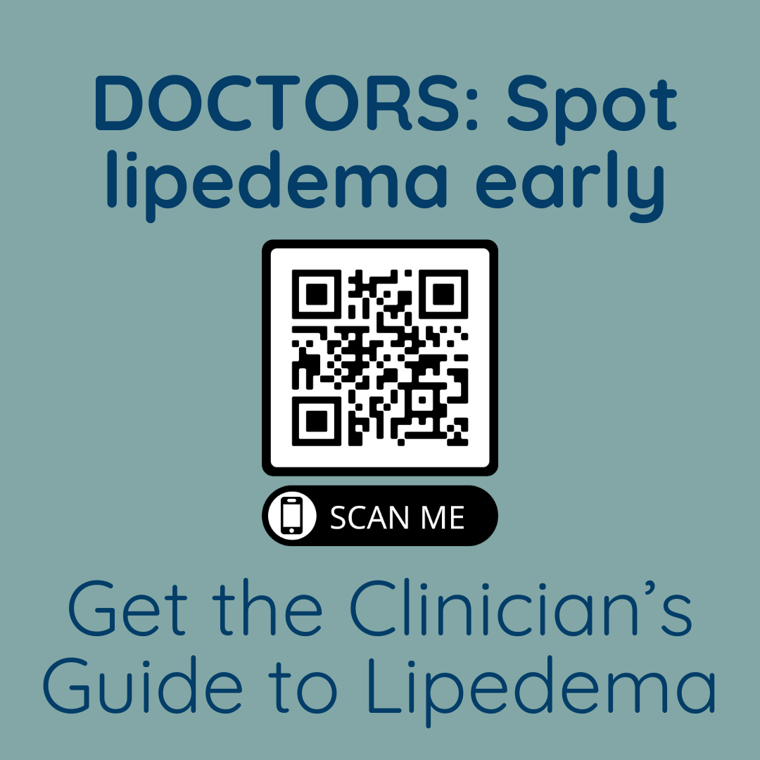
Lipedema affects 11% of women, yet it's frequently misdiagnosed or dismissed. Improve diagnoses, and support those living with #lipedema
#MedStudentTwitter #medstudents #MedEdChat #FutureDocs #medicalstudent #medicine #medicalschool #medstudent #doctor #medschool #premed #medical

3
5
510
7 Jun 2024
#MedEdChat will be on hiatus during the month of July. Our next #mededchat will be the first Thursday in August. Have a safe and fun summer!
1
3
369
6 Jun 2024
#MedEdChat is live tonight at 9 PM ET/NYC! Join us to discuss this approach to grading #medstudents! Is it better? Can it help with #CBME?
1
2
268
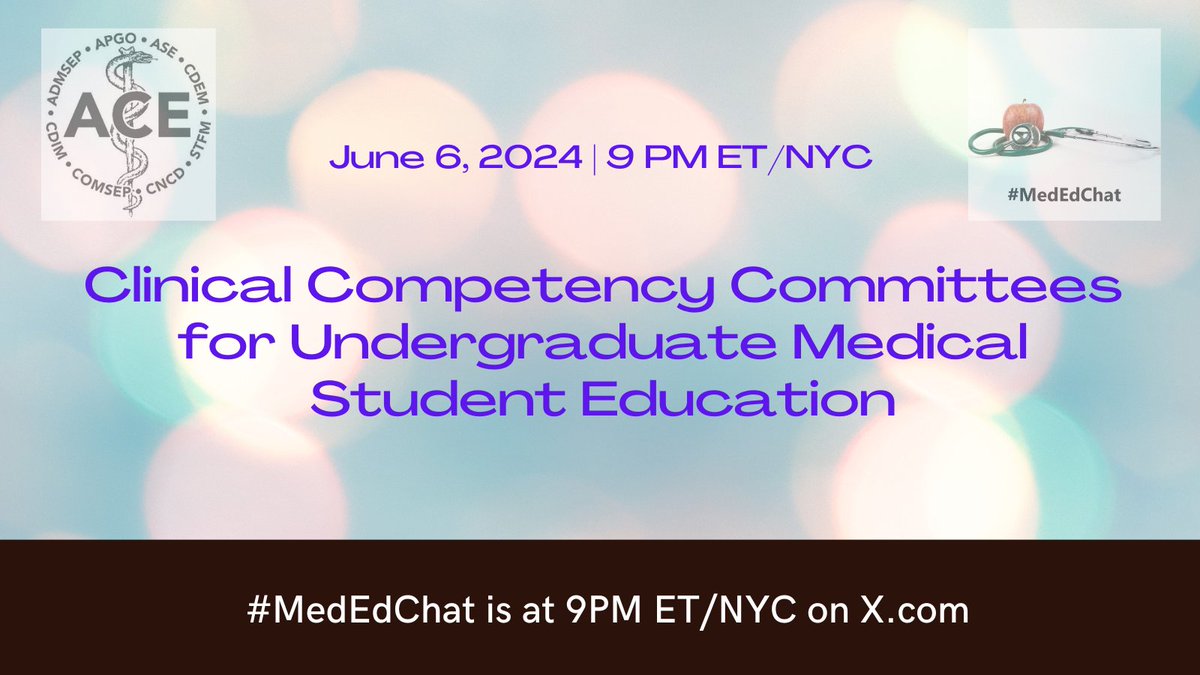
3 Jun 2024
Join #MedEdChat Thursday night to discuss clinical competency committees in #medstudent grading! #meded

1
3
564
31 May 2024
Kick off the summer with an exciting chat!
Join #MedEdChat on June 6th at 9 PM ET/NYC!
#meded #hmicommunity

2
3
505

3 May 2024
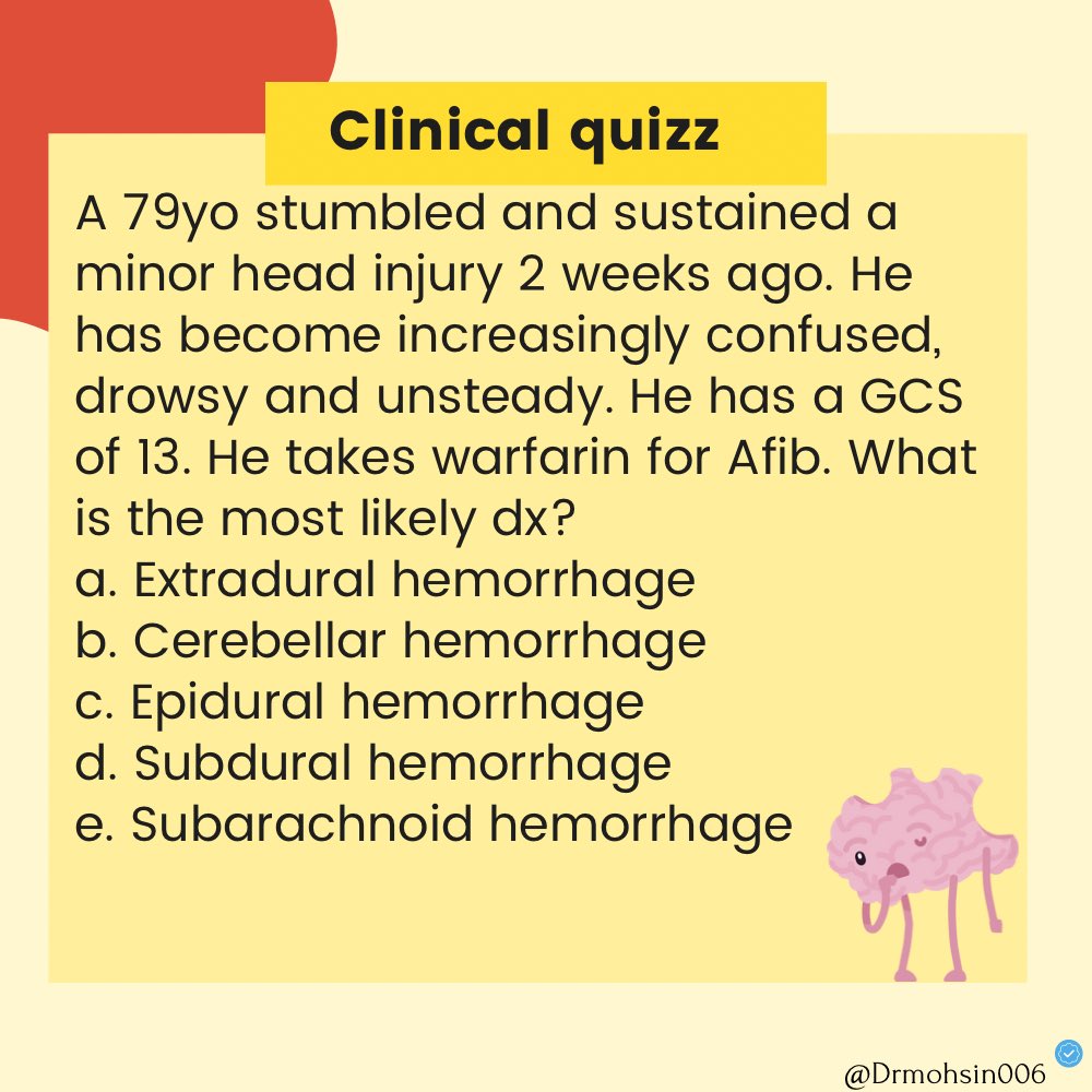
Are you up for a brainy challenge? 🤔 Test your wits with our Neurological Quiz and see if you can conquer the mysteries of the mind. Leave a comment to join the quest! 💡 🧠
#MedEdChat #MedEd #Neurology
5
2
8
969

3 May 2024
Medical students and doctors from less advantaged socio-economic backgrounds often face specific barriers when navigating education & training which may influence speciality career - and can be unfair. Paper here: sciencedirect.com/science/ar… #MedEd #MedEdChat #EDI
2
8
20
4,838

3 May 2024
We had an amazing chat this week on this topic in HPE-Global! @ASanchez_PS @hinterlandphpm @aminocte @timdyster
Maybe our class could host a #MedEdChat sometime?
3
152

3 May 2024
Some of my colleagues use #googlevoice on their personal phone to give as a separate number for #meded learners to call use. It’s not secure but it is “free”. Others use it? #MedEdChat
5
245

#mededchat T4 by having institutional policies including social media usage and developing ethical frameworks for these #meded
1
1
45