
The microscope is now data.
Digital pathology has moved past pilots. In 2025, FDA granted De Novo status to ArteraAI Prostate, software analyzing biopsy images for cancer prognosis [1]. The market is projected to grow from $1.46B to $2.75B by 2030 [2]. 🧬
The bottleneck is people: a 2026 report warns of a 16M diagnostic-staff shortfall by 2050 [3]. AI won’t replace pathologists; it will decide which labs scale and cut delays. 🔬
Leaders need scanners, validation, audit trails, and training not hype. ⚕️
🤔 What is the bigger risk: bias, broken workflows, or unequal access?
[1] [accessdata.fda.gov/SCRIPTs/c…)
[2] [marketsandmarkets.com/Market…](marketsandmarkets.com/Market…)
[3] [theguardian.com/society/2026…](theguardian.com/society/2026…)
#DigitalPathology #CancerAI
1
112

Jun 4
🧠 X Post: Pathologist-Free AI for Oral Cancer Detection & Grading
Dual-stage AI system for Pathologist-Free Tumor Detection and Subtyping in Oral Squamous Cell Carcinoma
📄 medRxiv (2026)
🔗 DOI: 10.1101/2025.06.05.25329090
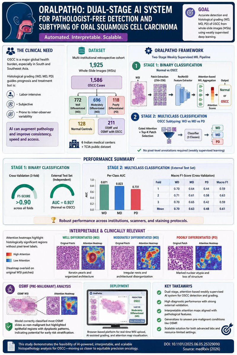
Oral squamous cell carcinoma (OSCC) remains one of the most common and deadly cancers in South and Southeast Asia, where access to expert pathology services is often limited. Histologic grading into well-differentiated (WD), moderately differentiated (MD), and poorly differentiated (PD) tumors is critical for prognosis and treatment decisions, but the process is labor-intensive and prone to inter-observer variability.
A new study introduces OralPatho, a dual-stage weakly supervised AI framework that performs automated tumor detection and histological grading directly from whole-slide pathology images (WSIs), without requiring pixel-level annotations.
The authors assembled one of the largest OSCC digital pathology datasets reported to date:
🔹 1,586 OSCC WSIs
🔹 772 WD tumors
🔹 696 MD tumors
🔹 118 PD tumors
🔹 Multi-institutional cohort spanning six Indian medical centers plus TCIA data
The pipeline operates in two stages:
1️⃣ Binary classifier (Normal vs OSCC) using attention-based Multiple Instance Learning (MIL)
2️⃣ Multiclass classifier assigning WD, MD, or PD grade using gated attention and Top-K patch selection
Each WSI is decomposed into 256×256 image patches, encoded via a ResNet50 backbone into 512-dimensional embeddings, then aggregated using attention mechanisms that mimic how pathologists focus on diagnostically relevant regions.
Performance was impressive:
✅ Binary detection F1 >0.90 across validation folds
✅ External-test AUC:
• WD: 0.871
• MD: 0.823
• PD: 0.731
✅ Robust performance despite differences in scanners, staining protocols, and institutions.
Perhaps the most interesting aspect is interpretability.
Attention heatmaps consistently localized classical pathological features:
🔬 Keratin pearls in WD tumors
🔬 Architectural disorganization in MD lesions
🔬 Nuclear atypia in PD tumors
Importantly, these explanations emerged without pixel-level annotations, providing transparency that is often missing in computational pathology workflows.
The team also tested the model on oral submucous fibrosis (OSMF), a premalignant condition never seen during training. Although most OSMF slides were classified as non-malignant, the model selectively highlighted epithelial regions showing subtle dysplastic patterns, suggesting potential future applications in early risk stratification and malignant transformation surveillance.
A browser-based deployment platform further enables real-time slide upload, AI-assisted grading, and visualization of attention maps, moving the system closer to clinical implementation in resource-limited settings.
This work demonstrates how weakly supervised foundation pathology approaches can extend beyond simple cancer detection toward explainable grading and pre-malignant lesion assessment—an important step toward scalable AI-assisted oral oncology.
#AIinMedicine #DigitalPathology #ComputationalPathology #OralCancer #OSCC #DeepLearning #Histopathology #CancerAI #PrecisionOncology #Pathology #MachineLearning #MedicalAI #OralPatho #WSI #MedTech
2
129

May 30
BREAKING: Tempus AI Launches Tempus Preview for Faster Cancer Insights $TEM unveiled Tempus Preview, a new AI-powered application designed to deliver rapid oncology decision support. Key Highlights: Provides preliminary biomarker insights within ~24 hours of tissue receipt
Bridges the gap before full genomic sequencing results are available
Initial focus on MSI-H, EGFR mutations, and FGFR fusions
Powered by Paige Predict AI trained on millions of slides
Aims to accelerate personalized cancer treatment planning
Expands Tempus’ AI-driven precision medicine tools in clinical workflows
#TEM #TempusAI #PrecisionOncology #CancerAI #Biomarkers #BREAKING
4
311

#AI is no longer a future concept in oncology- it is already reshaping research, diagnostics, clinical trials, and patient care.
Proud to be part of the growing European multidisciplinary community behind @ESAC_cancerAI working to advance responsible and clinically meaningful AI in cancer research. Reflecting on three amazing days in Milan for the 1st Annual ESAC Meeting- what a unique event and what a great time we had!
#AIinOncology #CancerAI #ESAC #HealthTech

1
2
15
704

[Nat Med] AITIC Trial - AI Triage in Breast Cancer Screening (Mar 2026)
Prospective non-inferiority trial (n=31,301) evaluating AI-based triage in mammography screening (Spain).
Design:
- Low-risk exams: AI reads autonomously (no radiologist needed)
- Remaining: double-read with AI decision support (Transpara, ScreenPoint Medical)
Results:
- Radiologist workload reduced by 63.6%
- Cancer detection rate increased from 6.3 to 7.3 per 1,000 ( 15.2%)
- Recall rate increased by 14.8%
AI triage offers a practical path to maintaining screening quality amid global radiologist shortages. The increased recall rate warrants monitoring.
nature.com/articles/s41591-0…
#BreastCancer #CancerAI #Mammography #RadiologyAI
2
126

🎤Please welcome to the stage @ElhamAzizi presenting on integrating AI & single-cell approaches to decode tumor micro-environments.
#IICDSymposium #CancerAI
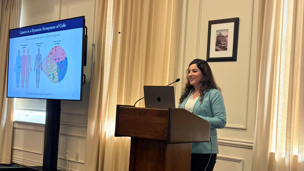
ALT Elham Azizi presents at the 2026 IICD Symposium.
1
1
14
539

Feb 1
Artificial intelligence agents in cancer research and oncology
nature.com/articles/s41568-0…
@NatureRevCancer
#AIagents #AIBio #CancerAI
1
4
1,545

21 Dec 2025
🔗 Apply & amplify impact: moffitt-cancer-center-career… moffitt-cancer-center-career… #AIinHealthcare #CancerAI #DataScienceJobs
1
2
64

1 Dec 2025
I had the opportunity to sit down with two exceptional leaders who are shaping the future of healthcare from very different, but deeply connected angles: #oncology innovation and industry-wide ecosystem growth.
First, @NaineshParikhMD of Moffitt Cancer Center (@MoffittNews) joins me for a powerful conversation on the future of cancer care: from budget strategy and governance to AI-driven clinical research and the bold question of whether we can move more cancers into chronic, survivable conditions.
Then, @richscarfo, President of @HLTHEVENT, shares what made #HLTH25 the most energized event yet and what’s ahead for the HLTH ecosystem: from new experience zones and national dinner series to global expansion across Europe.
📍Recorded live at HLTH25 in #LasVegas
Catch the episode now on your favorite platform: lnkd.in/eDNC8zFF
#DigitalHealth #HealthIT #Healthcare #AI #Innovation #GenAI #Data #ArtificialIntelligence #Cancer #CancerCures #CancerInnovation #CancerAI #CloudMatters #CPUMatters #CPU #GPU #APU #CEO #CMO #CIO #CTO #CMIO #ValueBasedCare #VBC #CCM #Strategy #Leadership #PublicPolicy #PodcastoftheYear #Growth #JustinBarnes #HITAdvisor #StrategicPlanning #HealthTech #HIThinkTank #SecretSauce #Predictions #KeyTrends #FutureofHealth #HLTHUSA

2
9
291

30 Oct 2025
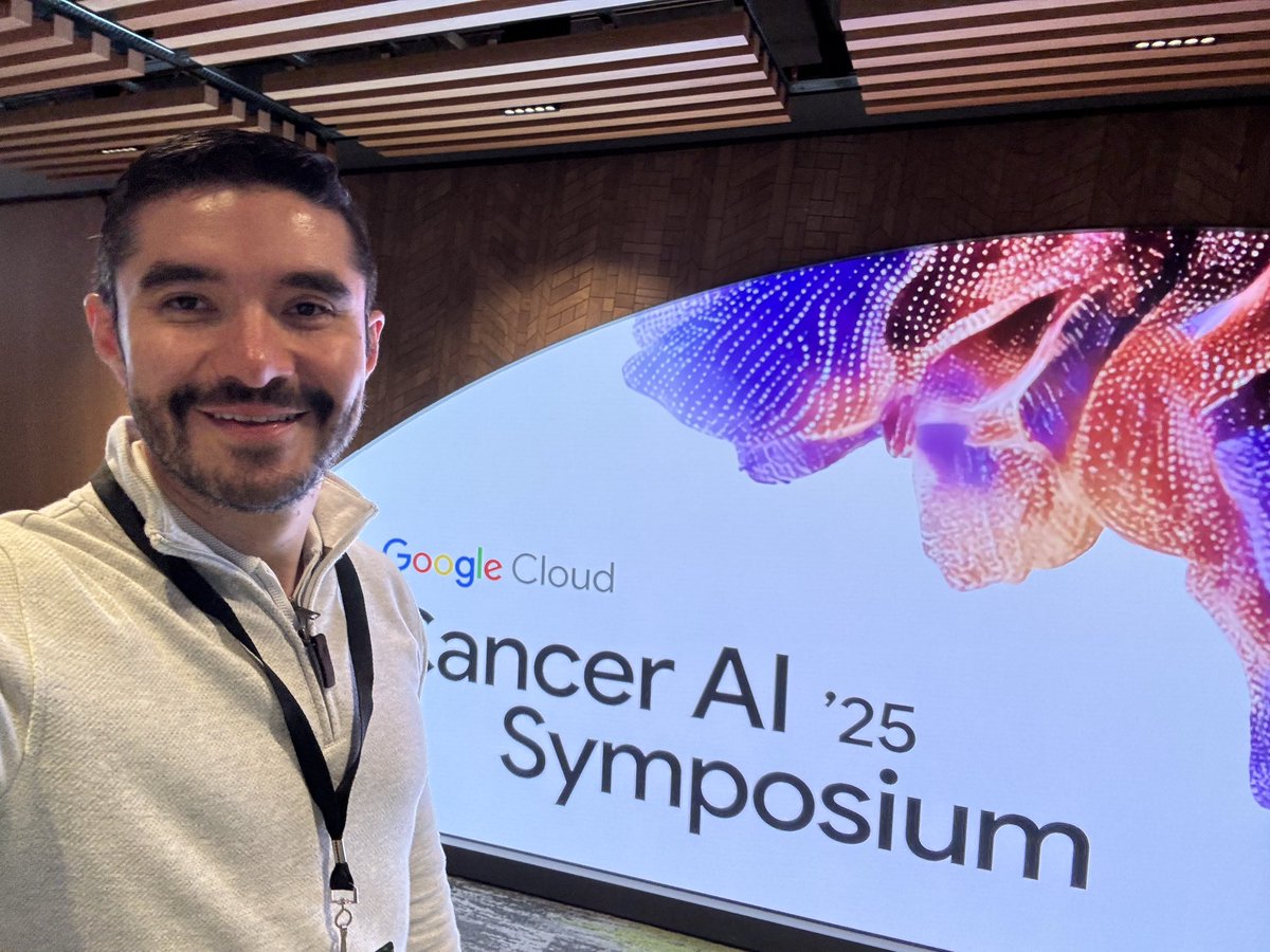
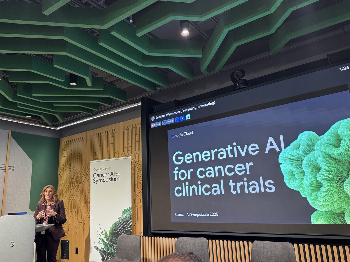
✨ At @Google HQ NYC for the Cancer AI Symposium 2025 — where oncology meets innovation.
Proud to represent @mystlukes and explore new #AI collaborations to bring cutting-edge cancer care to the Lehigh Valley.
Also thrilled that @MassiveBio is a sponsor — connecting with @skurnaz and incredible minds united to solve cancer.
#CancerAI #GoogleCloud #Oncology #AIinHealthcare #Innovation #StLukesProud
3
4
15
1,002
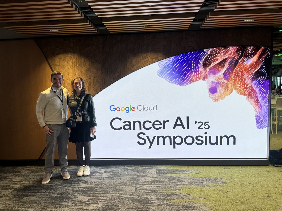
30 Oct 2025
Things just kicking off the Google Cloud AI in Cancer Symposium and incredible how AI is transforming cancer care. Excited about our growing efforts at Moffitt to advance AI-driven myeloma research. #CancerAI #OncologyInnovation
1
1
166

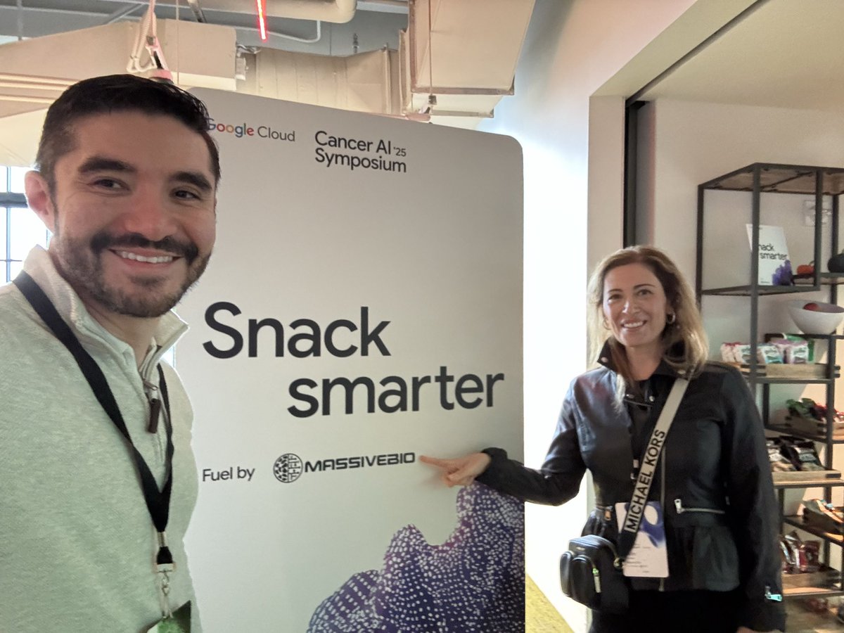
8 Sep 2025
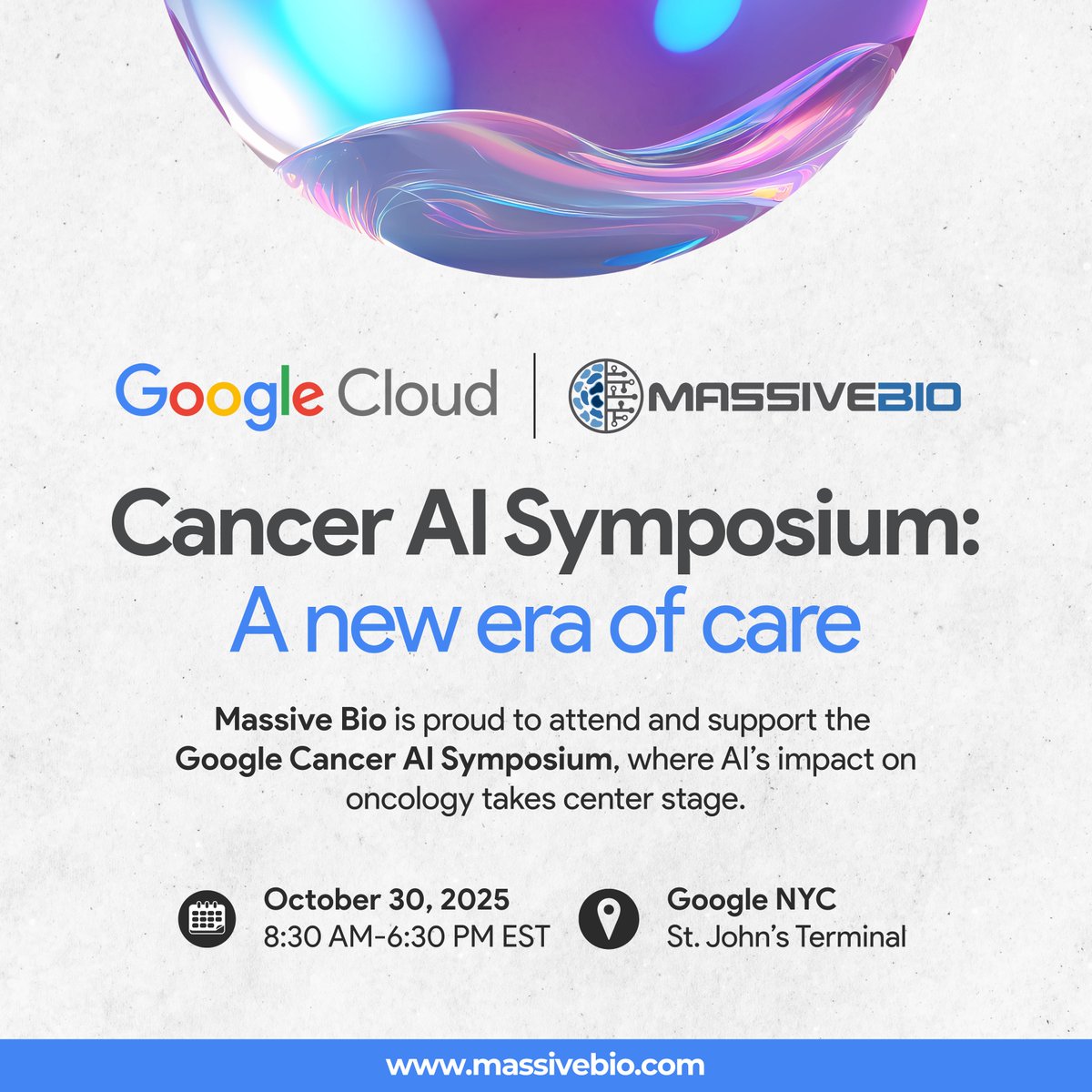
We proudly support the Cancer AI Symposium 2025, hosted by @googlecloud on Oct 30 in NYC. The event unites experts from oncology, pharma, and tech to discuss how AI is advancing cancer research and clinical trials.
Learn more: massivebio.com/massive-bio-a…
#CancerAI #Oncology #AI #ClinicalTrials #LifeSciences
1
6
789

2 Aug 2025
Can AI predict cancer recurrence before it happens?
📢 A new deep-learning model (RADAR CARE) predicts 1-year recurrence after curative surgery in early-stage NSCLC using multimodal clinical & radiologic data.
✅ 14,177 patients
✅ AUC: 0.854
✅ Sensitivity: 86%, Specificity: 71.3%
✅ Risk score independent of TNM stage
🧠 RADAR model uses transformer-based AI to process 64 clinical, molecular & imaging features, providing real-time recurrence risk with actionable thresholds:
•Low risk: RADAR < 0.3 (1-year recurrence <1%)
•Intermediate: 0.3–0.6
•High risk: >0.6 (recurrence ≥5%)
🔁 Four dynamic risk patterns suggest how to tailor follow-up & adjuvant therapy.
📌 Implication: Surveillance in early-stage NSCLC can now be personalized, moving beyond rigid TNM-based protocols.
📎 ascopubs.org/doi/full/10.120…
#LungCancer #NSCLC #Oncology #AIinHealthcare
#PrecisionOncology #DeepLearning #RecurrenceRisk
#CancerSurveillance #ThoracicOncology #RadOnc
#JCOPO #CancerAI #TransformerModel #MedicalAI
@JCO_ASCO @JCOGO_ASCO @ASCO @crisbergerot @osutcuoglu @Erman_Akkus @BbaharK @RyanNipp @brunolarvol @OncoAlert @oncodaily @OncoReporte @realbowtiedoc @ErulEnes @Xesiloglu @kbaskurtt

1
10
26
4,860

9 Jul 2025
Thrilled to collaborate with Mathew Garnett @Sanger and Parse Biosciences on the new Cancer Plasticity Atlas – a hugely exciting single cell genomics resource to decode tumor adaptation and resistance. 🔗 helmholtz-munich.de/en/newsr… #CancerAI #Plasticity #Atlas
1
15
77
6,335

1 Jul 2025
Emerging AI Approaches for Cancer Spatial Omics
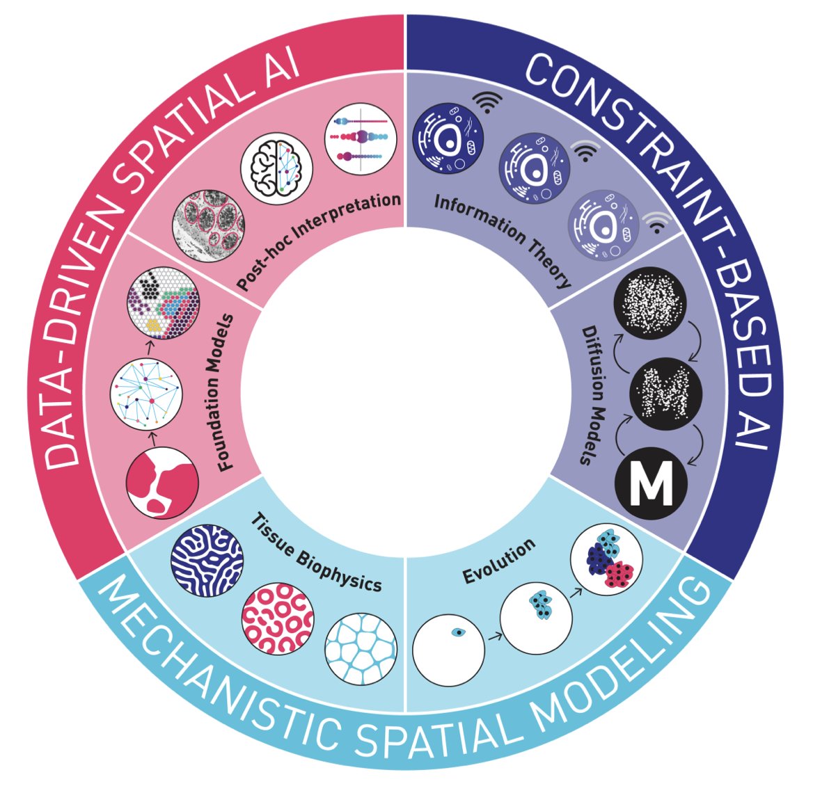
1.This paper introduces a comprehensive review of AI paradigms in cancer spatial omics, highlighting three key conceptual frameworks: data-driven spatial AI, constraint-based spatial AI, and mechanistic spatial modeling. The authors argue that interpretability and mechanistic grounding are crucial for clinical and biological utility.
2.Data-driven spatial AI is exemplified by foundation models trained on large histopathology or omics datasets. These models can perform tasks such as classification and annotation but often operate as black boxes, with limited biological interpretability.
3.Histopathology foundation models like DINOv2, Virchow, UNI, and GPFM are transformer-based and trained on millions of H&E slides. They excel in image tasks such as segmentation and diagnosis and are increasingly integrated with language models (e.g., CONCH, PathChat) for richer, multimodal interpretations.
4.Spatial omics foundation models are at an earlier stage, but tools like CellPLM and Nicheformer extend transformer methods to spatial transcriptomics, combining spatial and dissociated RNA data for tasks like label transfer and cell annotation.
5.Spatial proteomic models are emerging but face challenges in marker standardization. Recent efforts such as KRONOS demonstrate how tokenization strategies can improve model performance across heterogeneous datasets.
6.To improve interpretability, post-hoc techniques like SHAP, LIME, and attention mechanisms are employed. There is also growing interest in explainable structures like Functional Tissue Units (FTUs) and graph-based representations (e.g., SpaGFT) to ground features in biological contexts.
7.Constraint-based spatial AI introduces assumptions from biophysics and information theory. For example, models inspired by information bottleneck theory aim to compress spatial omics data while preserving biologically predictive signals.
8.Image diffusion models, adapted from generative AI (e.g., DALL-E, Stable Diffusion), are now applied to spatial omics. Methods like stDiff, DiffuST, and SpatialDiffusion use diffusion transformers and graph neural networks to impute missing spatial transcriptomics data and interpolate 3D structures.
9.Mechanistic spatial modeling offers the highest interpretability, aiming to infer biophysical processes directly. Physics-Informed Neural Networks (PINNs) and Kolmogorov–Arnold Networks (KANs) are proposed to learn equations from spatial data, capturing diffusion, tissue stiffness, and mechanical forces.
10.Cancer evolution can be tracked using spatial transcriptomics. Models like InferCNV, CalicoST, and transformer-based CNV callers are being adapted to spatial data to infer subclonal architecture and evolutionary pressures. There is potential in integrating expression, genotype, and morphology into unified AI models.
11.Data integration remains a challenge. Combining H&E, spatial transcriptomics, and proteomics requires careful standardization and alignment. Methods like SpatialGlue, COVET, and OmiCLIP aim to merge these modalities for domain identification and gene prediction.
12.The paper advocates for mechanism-driven data generation, particularly using mouse models. Mice allow dynamic and perturbative spatial omics data collection, crucial for training models like PINNs. Cross-species alignment tools (e.g., BrainAlign, Nicheformer) are in development to bridge human and mouse data.
13.Validation of spatial foundation models is still lacking. The authors argue that benchmarking should occur at multiple spatial scales and reflect the ability to distinguish fine-grained microenvironments. Mechanistic and constraint-based approaches may offer more interpretable and data-efficient alternatives.
14.The authors conclude that future progress hinges on curated cross-species datasets, consensus benchmarking tasks, and interpretable AI frameworks. These will enable better identification of functional tissue units and enhance clinical translation in cancer spatial omics.
📜Paper: arxiv.org/abs/2506.23857
#SpatialOmics #CancerAI #DeepLearning #ComputationalPathology #SpatialTranscriptomics #FoundationModels #ExplainableAI #TissueBiophysics #PINNs

2
6
700
1 Jul 2025
Emerging AI Approaches for Cancer Spatial Omics
1.This paper introduces a comprehensive review of AI paradigms in cancer spatial omics, highlighting three key conceptual frameworks: data-driven spatial AI, constraint-based spatial AI, and mechanistic spatial modeling. The authors argue that interpretability and mechanistic grounding are crucial for clinical and biological utility.
2.Data-driven spatial AI is exemplified by foundation models trained on large histopathology or omics datasets. These models can perform tasks such as classification and annotation but often operate as black boxes, with limited biological interpretability.
3.Histopathology foundation models like DINOv2, Virchow, UNI, and GPFM are transformer-based and trained on millions of H&E slides. They excel in image tasks such as segmentation and diagnosis and are increasingly integrated with language models (e.g., CONCH, PathChat) for richer, multimodal interpretations.
4.Spatial omics foundation models are at an earlier stage, but tools like CellPLM and Nicheformer extend transformer methods to spatial transcriptomics, combining spatial and dissociated RNA data for tasks like label transfer and cell annotation.
5.Spatial proteomic models are emerging but face challenges in marker standardization. Recent efforts such as KRONOS demonstrate how tokenization strategies can improve model performance across heterogeneous datasets.
6.To improve interpretability, post-hoc techniques like SHAP, LIME, and attention mechanisms are employed. There is also growing interest in explainable structures like Functional Tissue Units (FTUs) and graph-based representations (e.g., SpaGFT) to ground features in biological contexts.
7.Constraint-based spatial AI introduces assumptions from biophysics and information theory. For example, models inspired by information bottleneck theory aim to compress spatial omics data while preserving biologically predictive signals.
8.Image diffusion models, adapted from generative AI (e.g., DALL-E, Stable Diffusion), are now applied to spatial omics. Methods like stDiff, DiffuST, and SpatialDiffusion use diffusion transformers and graph neural networks to impute missing spatial transcriptomics data and interpolate 3D structures.
9.Mechanistic spatial modeling offers the highest interpretability, aiming to infer biophysical processes directly. Physics-Informed Neural Networks (PINNs) and Kolmogorov–Arnold Networks (KANs) are proposed to learn equations from spatial data, capturing diffusion, tissue stiffness, and mechanical forces.
10.Cancer evolution can be tracked using spatial transcriptomics. Models like InferCNV, CalicoST, and transformer-based CNV callers are being adapted to spatial data to infer subclonal architecture and evolutionary pressures. There is potential in integrating expression, genotype, and morphology into unified AI models.
11.Data integration remains a challenge. Combining H&E, spatial transcriptomics, and proteomics requires careful standardization and alignment. Methods like SpatialGlue, COVET, and OmiCLIP aim to merge these modalities for domain identification and gene prediction.
12.The paper advocates for mechanism-driven data generation, particularly using mouse models. Mice allow dynamic and perturbative spatial omics data collection, crucial for training models like PINNs. Cross-species alignment tools (e.g., BrainAlign, Nicheformer) are in development to bridge human and mouse data.
13.Validation of spatial foundation models is still lacking. The authors argue that benchmarking should occur at multiple spatial scales and reflect the ability to distinguish fine-grained microenvironments. Mechanistic and constraint-based approaches may offer more interpretable and data-efficient alternatives.
14.The authors conclude that future progress hinges on curated cross-species datasets, consensus benchmarking tasks, and interpretable AI frameworks. These will enable better identification of functional tissue units and enhance clinical translation in cancer spatial omics.
📜Paper: arxiv.org/abs/2506.23857
#SpatialOmics #CancerAI #DeepLearning #ComputationalPathology #SpatialTranscriptomics #FoundationModels #ExplainableAI #TissueBiophysics #PINNs
2
7
1,021
9 May 2025
AI-powered integration of multi-source data for TAA discovery to accelerate ADC and TCE drug development (I): TAA Target Identification and Prioritization
1.This paper presents a GraphRAG-enhanced GPT-4 framework for discovering and prioritizing tumor-associated antigens (TAAs), aiming to improve the precision and efficiency of antibody-drug conjugate (ADC) and T-cell engager (TCE) development.
2.The method integrates diverse data sources—bulk and single-cell RNA-seq, spatial transcriptomics, proteomics, and biomedical literature—creating a unified, scalable, and data-agnostic platform for TAA target triage.
3.A major innovation is the use of a safety score derived from z-scored gene expression across normal tissues, which helps eliminate TAAs with high off-tumor risks and ensures tumor selectivity.
4.The GraphRAG model constructs a biological knowledge graph from PubMed data, continuously updated with new findings and refined through a feedback loop to improve its relevance and accuracy in TAA identification.
5.To demonstrate utility, the authors compare the base GPT-4 and their RAG-enhanced model on the evaluation of MUC1 and TACSTD2 in NSCLC. The enhanced model produced more nuanced, evidence-backed, and clinically aligned insights.
6.MUC1, with a higher safety score and clearer functional role in immune evasion and metastasis, was prioritized by the model as a superior ADC/TCE target, consistent with current therapeutic development trends.
7.The framework supports an interactive platform where users can submit gene and cancer type prompts and receive interpretable, evidence-integrated reports, streamlining the discovery-to-evaluation pipeline.
8.This study highlights how prompt engineering and domain-specific augmentation significantly improve LLM outputs, enabling better biomedical knowledge synthesis and clinical relevance.
9.Beyond current functionality, the authors envision incorporating additional modalities (e.g., digital pathology images), extending the system's use to broader cancer types and enabling deeper multimodal TAA profiling.
10.The authors emphasize the importance of experimental validation and suggest future directions including combinatorial therapy modeling and expanded biomarker discovery.
11.Overall, this work demonstrates how domain-enhanced large language models, paired with multi-omics integration, can meaningfully accelerate cancer target discovery while improving safety, interpretability, and translational relevance.
📜Paper: doi.org/10.1101/2025.05.06.6… #CancerAI #ADC #TCE #TumorAntigen #GraphRAG #OmicsIntegration #DrugDiscovery #LLM #Bioinformatics #Oncology
15
942
9 May 2025
AI-powered integration of multi-source data for TAA discovery to accelerate ADC and TCE drug development (I): TAA Target Identification and Prioritization
1.This paper presents a GraphRAG-enhanced GPT-4 framework for discovering and prioritizing tumor-associated antigens (TAAs), aiming to improve the precision and efficiency of antibody-drug conjugate (ADC) and T-cell engager (TCE) development.
2.The method integrates diverse data sources—bulk and single-cell RNA-seq, spatial transcriptomics, proteomics, and biomedical literature—creating a unified, scalable, and data-agnostic platform for TAA target triage.
3.A major innovation is the use of a safety score derived from z-scored gene expression across normal tissues, which helps eliminate TAAs with high off-tumor risks and ensures tumor selectivity.
4.The GraphRAG model constructs a biological knowledge graph from PubMed data, continuously updated with new findings and refined through a feedback loop to improve its relevance and accuracy in TAA identification.
5.To demonstrate utility, the authors compare the base GPT-4 and their RAG-enhanced model on the evaluation of MUC1 and TACSTD2 in NSCLC. The enhanced model produced more nuanced, evidence-backed, and clinically aligned insights.
6.MUC1, with a higher safety score and clearer functional role in immune evasion and metastasis, was prioritized by the model as a superior ADC/TCE target, consistent with current therapeutic development trends.
7.The framework supports an interactive platform where users can submit gene and cancer type prompts and receive interpretable, evidence-integrated reports, streamlining the discovery-to-evaluation pipeline.
8.This study highlights how prompt engineering and domain-specific augmentation significantly improve LLM outputs, enabling better biomedical knowledge synthesis and clinical relevance.
9.Beyond current functionality, the authors envision incorporating additional modalities (e.g., digital pathology images), extending the system's use to broader cancer types and enabling deeper multimodal TAA profiling.
10.The authors emphasize the importance of experimental validation and suggest future directions including combinatorial therapy modeling and expanded biomarker discovery.
11.Overall, this work demonstrates how domain-enhanced large language models, paired with multi-omics integration, can meaningfully accelerate cancer target discovery while improving safety, interpretability, and translational relevance.
📜Paper: doi.org/10.1101/2025.05.06.6…
#CancerAI #ADC #TCE #TumorAntigen #GraphRAG #OmicsIntegration #DrugDiscovery #LLM #Bioinformatics #Oncology

2
522

5 May 2025

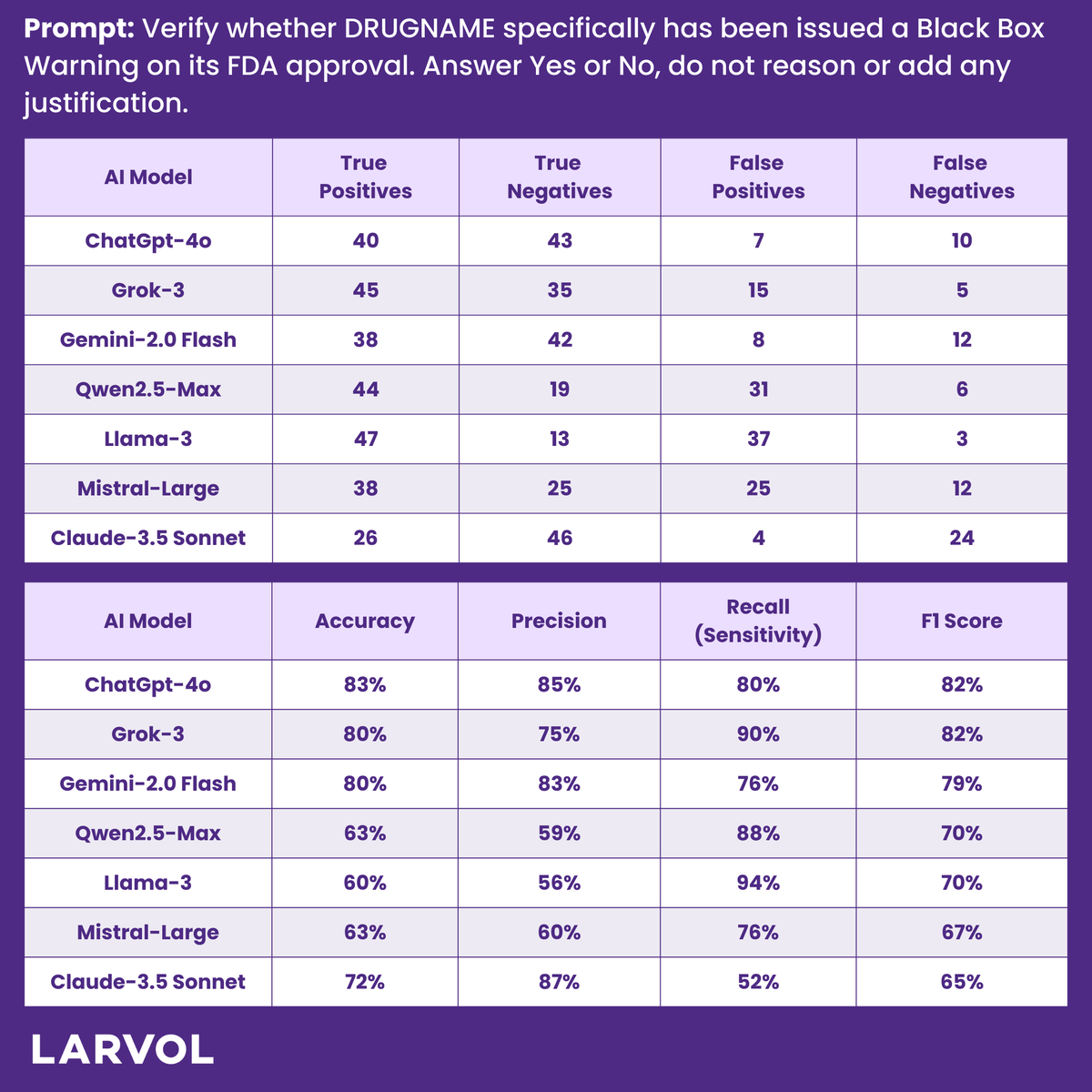
Last time, we challenged AI systems to identify specific effects in Black Box Warnings. This time, we raised the stakes: Can AI accurately determine whether a drug has any FDA Black Box Warning at all?
Here’s how we stress-tested the #AIS:
1. Expert-Crafted Prompts – Multiple variations to ensure robustness.
2. AI Battle-Testing – Identical prompts across leading AI platforms for a fair comparison.
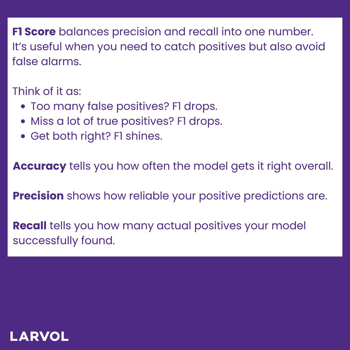
3. Performance Ranking – Scored using F1 (precision recall) to minimize false positives while capturing true positives.
4. Iterative Refinement – Optimized until we found the winning approach.
The results? See below!
Let's dive deeper into our methodology & results in the comments.
#LARVOL #CancerResearch #Oncology #CancerData #ClinicalTrials #ChatGPT #Grok #Gemini #Qwen #Llama #MistralAI #Claude | @Larvol | @brunolarvol | @one_interrobang | @Davdwlkerson | @SuyogCancer | @DrBonillaOnc | @MedinDarko

5
5
310

25 Apr 2025
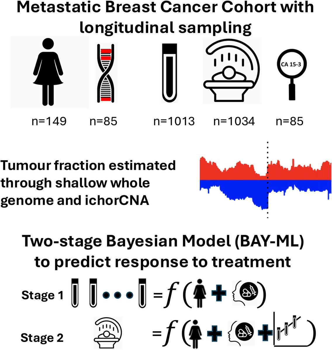
🧬💻A large-scale retrospective study in metastatic breast cancer patients using circulating tumour DNA and machine learning to predict treatment outcome and progression-free survival
👉 buff.ly/jWEEbZi
@stephensammut #BCSM #CancerAI #ctDNA
6
12
387